Superficial Inguinal Lymph Nodes
a. Superficial Inguinal Lymph Nodes of Male Dogs
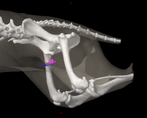
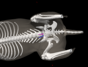
The superficial inguinal lymph nodes of male dogs (Figures 31, 32: 4) form a group of 1 to 3 (usually 2) lymph nodes on each side, which lie in the very fatty subcutaneous tissue on the dorsolateral edge of the penis, between the penis and the ventral abdominal wall, 0.5 to 1.0 cm cranial from the spermatic cord. The lymph nodes lie along the cranial border of the medial surface of the M. gracilis, and ventral to the aponeurosis of the M. obliquus externus abdominis.
A different number of lymph nodes on the right and left sides were often observed.
The size of the lymph nodes is highly variable: they were 5 mm to 6.8 cm in length, 2.5 cm in width, and 1.5 cm in thickness.
The position of the superficial inguinal lymph node(s) in relation to the external pudendal artery and vein, which run through the fat surrounding the lymph nodes, is as follows: if there is 1 lymph node, it is located medial to the vessels, between the vessels and the ventral abdominal wall, and protrudes cranially and caudally over the vessels. If there are 2 lymph nodes, 1 is almost always located on the cranial border of the vessels and the second on the caudal border of the vessels, so that the vessels pass (in the craniomedial direction) between both lymph nodes. More infrequently, 1 lymph node may be medial to the vein and the other caudal to it. If there are 3 lymph nodes, 1 is cranial to the external pudendal artery and vein, and the other 2 lie immediately adjacent to one another, caudal to the vessels. One of the 2 caudal lymph nodes may be located medial to the spermatic cord. The cranial lymph node may be located more deeply, while the caudal lymph nodes are located more superficially.
The absolute weight of the lymph nodes on both sides varied between 0.02 g and 23.23 g, the relative weight between 0.0003% and 0.0719%.
Efferent drainage
The superficial inguinal lymph nodes on one side are usually connected to one another by efferent vessels (Figure 31: 7). In addition, the efferent vessels of the individual lymph nodes will merge to form 1 to 2 larger vessels, which run caudodorsally over the medial surface of the spermatic cord, accompanying the external pudendal artery and vein, eventually entering the abdominal cavity along the external iliac artery and vein, and finally draining into the medial iliac lymph node (Figures 31: 1; 27: 11). If a deep inguinal lymph node (Figure 27: 8) is present, some or all the efferent vessels enter this lymph node.
b. Superficial inguinal lymph nodes of female dogs (Lnn. supramammarii, mammary lymph nodes, Figure 30: 1)
Of 7 bitches examined, 4 had only 1 superficial inguinal lymph node on each side, 2 had 2 lymph nodes on each side, and 1 had 2 lymph nodes on the right side and 1 lymph node on the left side.
In large dogs, the lymph nodes are up to 1 to 2 cm long, up to 1 cm wide, and up to 0.5 cm thick.
Depending on the size of the dog, the lymph nodes on each side lie 2 to 4 cm cranial to the pecten ossis pubis and 1 to 1.25 cm lateral to the linea alba, and are therefore located between the ventral abdominal wall, the mammary tissue, and the skin of the ventral abdominal wall, close to the region where the skin of the ventral abdominal wall becomes the skin of the medial surface of the thigh. The external pudendal artery and vein, running in the cranial direction, usually lie along the lateral border of the lymph node. If there are two lymph nodes on one or both sides, they are then either located immediately adjacent to one another, or one is lateral and one is medial to the aforementioned vessels. The lateral lymph node usually then extends to the lateral edge of the mammary tissue and often protrudes slightly beyond it.
The absolute weight of the lymph nodes on both sides varied between 0.15 g and 1.16 g, and the relative weight between 0.0012% and 0.0178%.
efferent drainage
The efferent vessels in the female dog behave essentially the same as in the male. From each lymph node, they merge to form 1 to 2 larger vessels that enter the abdominal cavity with the external pudendal artery and vein (Figure 27: 11) and then accompany the external iliac artery and vein to drain to the medial iliac lymph nodes (Figure 27: 42).
If a deep inguinal lymph node is present (Figure 27: 8), then all or some of the efferent vessels open into it (Figure 27: 11′).
Afferent drainage in both males and females
The superficial inguinal lymph nodes drain lymph vessels from the skin of the ventral half of the portion of the abdominal wall located caudally from a transverse plane at the level of the last rib, including the lymph vessels of the skin of the prepuce and scrotum, the skin of the mammary gland, the skin of the caudal part of the pelvis, and of the tail, the lateral and medial sides of the thigh, the medial side and the cranial half of the lateral side of the stifle joint, the medial side and the cranial half of the lateral side of the lower leg (including the anterior border), the medial, the flexor, and the extensor sides of the tarsus, metatarsus, and digits, the lymph vessels of the abdominal skin muscles, the lymph vessels of the vulva, clitoris, and mammary glands, the scrotum, prepuce, and penis (including glans), and finally the lymph vessels of the male urethra.
Clinical Notes
The superficial inguinal lymph nodes on each side are usually connected to one another by efferent vessels.


