
Figure 15: Lymph Vessels of the Salivary Glands of the Dog
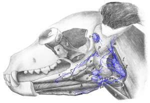
Figure 15: a M. mylohyoideus (retracted); b M. geniohyoideus; c M. genioglossus; d M. styloglossus; e M. hyoglossus; f aboral part of M. digastricus (cut off); g M. pterygoideus; h eye muscles; i lacrimal gland; k zygomatic gland; l M. sternomastoideus and cleidomastoideus (a piece cut out of both to allow the medial retropharyngeal lymph node [t] to be exposed); m pharyngeal muscles; n M. hyothyroideus; o M. sternohyoideus; p parotid gland and q submaxillary (mandibular) gland (a piece cut out of both glands); r caudal part of sublingual gland (glandula sublingualis grandicanalaris) and r’ rostral part of sublingual gland (glandula sublingualis parvicanalaris); s parotid lymph node; t medial retropharyngeal lymph node; u lateral retropharyngeal lymph node; v, v1, v2, v3 mandibular lymph nodes. 1 lymph vessel of the sublingual gland (glandula sublingualis parvicanalaris) running to a mandibular lymph node (cut off). Source: Dr. Hermann Baum (1918). (This work is in the public domain).
