
Figure 21: Left Side of the Dog’s Heart with Injected Lymph Vessels
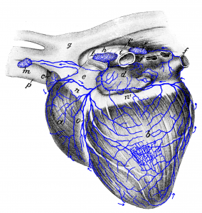 Figure 21: a right ventricle; b left ventricle; c right atrium (auricle); d left atrium; e pulmonary artery (cut off); f pulmonary veins (cut off); g aorta; h end of the trachea; i left and i’ right main bronchus; k middle tracheobronchial lymph node; l left tracheobronchial lymph node; m cranial mediastinal lymph node; n, n’ coronary sulcus; o left longitudinal sulcus; p cranial vena cava. Source: Dr. Hermann Baum (1918). (This work is in the public domain).
