
Figure 18: Right Side of the Thoracic Cavity of the Dog
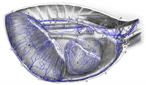 Figure 18: After removal of right costal wall and right lung. a left ventricle; b right ventricle; c right atrium; d, d’ coronary sulcus; e right longitudinal sulcus; f caudal vena cava; g cranial vena cava; h azygos vein; i external jugular vein; i’ internal jugular vein; k internal mammary artery and vein; l right subclavian artery; m right axillary artery and vein; n right costocervical vein; o right vertebral artery; p right common carotid artery; q aorta; r trachea; s right main bronchus; s’ right eparterial bronchus; t esophagus; u M. longus colli; v left M. transversus thoracis; w costal part, w1 lumbar part, and w2 central tendon of diaphragm; x sternum; y, y’ dorsal and ventral piece of 1st rib; z right M. transversus thoracis (cut off). 1 middle tracheobronchial lymph node; 2 right tracheobronchial lymph node; 3, 31, 32, 33, 34, 35 cranial mediastinal lymph nodes; 4 lymph vessels of the esophagus entering the abdominal cavity; 5, 5 lymph vessels of the esophagus turning towards its dorsal edge and then to the left and entering the left tracheobronchial lymph node; 6 sternal lymph node; 7 lymph vessels opening into the gastric lymph node, splenic lymph node, hepatic lymph node or cranial lumbar aortic lymph node; 8, 8 lymph vessels running to cranial lumbar aortic lymph node; 9 intercostal lymph node; 10 efferent vessel of a left cranial mediastinal lymph node; 11 thoracic duct; 12 right tracheal duct. Source: Dr. Hermann Baum (1918). (This work is in the public domain).
