Retropharyngeal Lymph Centre
The retropharyngeal lymph nodes include a medial retropharyngeal lymph node (Ln. retropharyngeus medialis) and, occasionally, a lateral retropharyngeal lymph node (Ln. retropharyngeus lateralis).
a. Medial retropharyngeal lymph node
The medial retropharyngeal lymph node (Figure 16: a) is an elongated, flattened lymph node on each side of the head, 1.5 to 8.0 cm long, 0.5 to 1.0 cm wide, and 0.5 to 1.0 cm thick, with a lateral and medial side, two blunted margins, and two blunted ends. It lies dorsally on the lateral wall of the pharynx (Figure 16: 12), immediately caudal to the M. digastricus (Figure 16: 14), and ventromedial to the free edge of the atlas wing, enveloped in fatty connective tissue. The caudal end usually extends to the caudal border of the M. cricopharyngeus. The rostral half of the medial retropharyngeal lymph node is located medially to the mandibular gland, and the caudal half is located medially to the M. sternomastoideus and M. cleidomastoideus, as well as to the ventral branch of the accessory nerve which enters the M. sternomastoideus. The medial retropharyngeal lymph node is located laterally to the M. longus capitis (Figure 16: 13) and the pharyngeal muscles (M. cricopharyngeus and thyropharyngeus) (Figure 16: 12), as well as the vessels and nerves associated with these muscles (common carotid artery, origin of occipital and internal carotid arteries, pharyngeal and cranial laryngeal branches of vagus nerve, hypoglossal nerve, and sympathetic trunk including the depressor nerve).
In 10 of 47 cases, there were either 2 medial retropharyngeal lymph nodes on both sides or on one side (Figures 8; 16: a, a’); in 2 cases on both sides, in 2 cases only on the left side, and in 6 cases only on the right side. When 2 lymph nodes are present, they are observed to be stacked, with the dorsal lymph node (Figure 16: a’) always being considerably smaller than the ventral lymph node, rarely more than 1.0 cm in length, even in large dogs. The smaller dorsal lymph node is located on the dorsomedial border of the larger ventral lymph node and does not protrude caudally beyond it.
If the dorsal lymph node is located towards the caudal end of the main ventral node, it may be mistaken for a cranial cervical lymph node; however, on examination of the drainage area, I have shown that this smaller dorsal lymph node is indeed a second medial retropharyngeal lymph node (see cranial cervical lymph node). In rare cases, the main medial retropharyngeal lymph node does not quite extend to the M. digastricus. Conversely, if the lymph node is unusually large, it extends to the cranial end of the thyroid gland(s) – in 1 case I examined, the main lymph node reached the dorsomedial margin of the thyroid gland (Figure 10: a). In these cases, the caudal end of the lymph node covers the cranial portion of the trachea.
The absolute weight of the lymph node(s) on both sides varied between 0.32 and 21.84 g, and the relative weight between 0.0135% and 0.0691%
Afferent drainage
The medial retropharyngeal lymph node(s) receive afferent lymph vessels from the skin of the pinna, lymph vessels from the M. omotransversarius, brachiocephalicus, sternomastoideus, rhomboideus, splenius, longissimus capitis, semispinalis capitis, longus colli, longus capitis and rectus capitis ventralis, the Mm. recti capitis dorsales and Mm. obliqui capitis, the M. sternohyoideus and sternothyroideus, as well as from various head bones (parietal, occipital, temporal, sphenoidal, palatine and mandible), the 1st and 2nd cervical vertebrae, various head muscles (M. masseter, digastricus, pterygoideus, and hyoid muscles), the tongue, the gingiva, the mucous membrane of the free floor of the oral cavity, the hard and soft palate, the tonsil, the parotid, mandibular and sublingual glands, the pharynx and esophagus, the nasal cavity, larynx, thyroid, trachea (initial part) and ear (muscles and labyrinth), the temporomandibular joint and the nervous system, as well as the efferent vessels from the mandibular, parotid and lateral retropharyngeal lymph nodes. If the medial retropharyngeal lymph node is double (Figures 8; 16: a, a’), then the afferent vessels can drain into both lymph nodes.
As would be expected, if only 1 or 2 afferent lymph vessels are injected, they usually only drain into 1 of the 2 lymph nodes. When a second medial retropharyngeal lymph node is present, all or at least most of the afferent lymph vessels from the neck muscles mentioned above drain into it, reaffirming that this second node is a retropharyngeal lymph node and not a cranial cervical lymph node.
efferent drainage
Many small efferent vessels emerge from the medial retropharyngeal lymph node, however, these immediately combine to form 3 to 5 larger efferent vessels. These vessels then combine to form the left and right tracheal duct (Figures 2, 4, 6, 8, 9, 10, 11, 12 and 16: a). One or 2 of these efferent vessels usually drain into a cranial cervical lymph node, if one is present (Figures 6 and 9: a, b). If there are 2 medial retropharyngeal lymph nodes on top of one another, the efferent vessels of the main lymph node behave as described above. In this case, usually 1 efferent vessel arises from the smaller dorsal lymph node, and either quickly merges with the efferent vessels of the larger ventral lymph node or converges with them to form the tracheal duct (Figure 16: a’).
b. Lateral Retropharyngeal Lymph Node
The lateral retropharyngeal lymph node (Figure 14: s” and 15: u) is a small, rarely double lymph node that is found in only about a third of all cases (see below). This lymph node lies in the fatty connective tissue ventromedial to the edge of the atlas, on the dorsal border of the mandibular gland, and along the terminal tendon of the M. sternomastoideus or on the origin of the M. digastricus near the cartilaginous auditory canal (Cartilago tubae auditivae). The lymph node is either completely or partially covered by the parotid gland. In the latter case, the lymph node can be observed to protrude slightly beyond the caudal border of the parotid gland (Figures 14: s”; 15: u). In 39 cases I examined in more detail, 27 dogs had no lateral retropharyngeal lymph node on either side. Of the remaining 12 cases, 8 dogs had a single lymph node on each side, 2 dogs had 1 lymph node on the right and 2 on the left, 1 dog had 1 lymph node on the left and 2 on the right, and 1 dog had 2 lymph nodes on the right 3 on the left. Most of the lymph nodes were 0.50 to 0.75 cm in size.
Afferent drainage
The lateral retropharyngeal lymph node(s) receive afferent lymph vessels from muscles located on the 1st and 2nd cervical vertebrae (M. cleidocervicalis, sternomastoideus, splenius, obliqui capitis, semispinalis capitis, dorsal recti, longissimus capitis, and the muscles of the cervical skin), as well as lymph vessels from the caudal ear muscles and the pinna, and the efferent vessels of the mandibular and parotid lymph nodes. Notably, the lymph vessels draining the skin were never observed to flow into the node.
Based on the drainage area described above, this lymph node evidently corresponds to the lateral retropharyngeal lymph node in cattle, and thus is not a parotid lymph node as reported by Merzdorf [24].
efferent drainage
One to 2 efferent vessels have been observed to emerge from each lateral retropharyngeal lymph node which drain into the medial retropharyngeal lymph node(s). The lateral retropharyngeal lymph nodes may also receive lymph from the medial retropharyngeal lymph node via retrograde flow.
Clinical Notes
Medial retropharyngeal lymph nodes
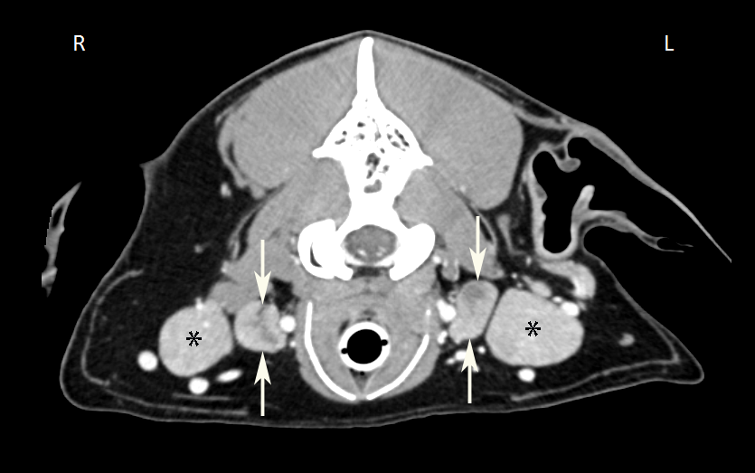
The deep location of the medial retropharyngeal lymph nodes does not allow fine needle aspiration without imaging guidance unless the lymph nodes are markedly enlarged; usually, ultrasound is used to guide the needle tip into the lymph node.