
Figure 28: Lymph Vessels of the Liver of the Dog
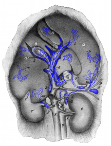
Figure 28: The animal is lying on its back; the visceral surface of the liver is shown. 1 left hepatic lymph nodes; 2 right hepatic lymph node; 3, 3′ cranial lumbar aortic lymph nodes; 4, 4, 4 subserosal lymph vessels starting to run deep; 5, 5 subserosal lymph vessels running subserosally to the hepatic lymph nodes; 6, 6 deep lymph vessels of the liver; 7, 7′ lymph vessels running with the terminal part of the esophagus; 8, 8‘splenic lymph nodes; 9, 9′ subserosal lymph vessels arising from the parietal surface of the liver; 10, 10′ subserosal lymph vessels from the visceral surface of the liver running to cranial lumbar aortic lymph nodes (3, 3′). a, a’ liver; b gallbladder; c left and c’ right kidney; d, d’ adrenal glands; e portal vein; f gastrosplenic vein; g aorta; h renal artery; i A. lumboabdominalis; k caudal vena cava; l V. lumboabdominalis; m renal vein; n esophagus (cut-off). Source: Dr. Hermann Baum (1918). (This work is in the public domain).
