
Figure 14: Lymph Vessels of the Tongue, Tongue Muscles, Soft Palate, and Larynx of the Dog
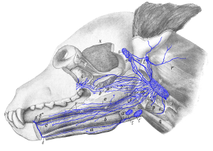
Figure 14: a, a’ M. mylohyoideus (largely retracted); b M. geniohyoideus; c M. genioglossus; d M. hyoglossus; e M. styloglossus; f M. pterygoideus; g eye muscles; h lacrimal gland; i zygomatic gland; k temporal muscles; l aboral end of the M. digastricus; m M. keratopharyngeus; n M. thyropharyngeus; o M. cricopharyngeus; p M. hyothyroideus; q M. sternohyoideus; r M. splenius; s, s’ parotid lymph nodes; s” lateral retropharyngeal lymph node; t, t’ mandibular lymph nodes; u medial retropharyngeal lymph node. 1, 1′ lymph vessels from hard palate and soft palate; 1″ lymph vessels of the zygomatic gland, joining 1′ labeled lymph vessels; 2, 2′ lymph vessels from the soft palate or 2′ lymph vessel from the fold surrounding the tonsillar sinus; 3, 3 lymph vessels from the tonsil; 4, 4′, 4″ lymph vessels from the soft palate, the tonsil, the base of the tongue and from the mucous membrane of the cavity of the pharynx, which initially run between the mucous membrane and the musculature; 5, 5 lymph vessels from the tongue, which perforate the M. hyoglossus; 6, 6′, 6″ lymph vessels of the tip of the tongue; 7, 7′ and 8 lymph vessels of the body of the tongue; 9, 9′ lymph vessels from the larynx. Source: Dr. Hermann Baum (1918). (This work is in the public domain).
