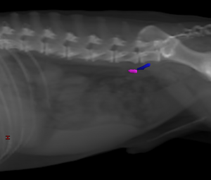
Hypogastric (Internal Iliac) Lymph Nodes
The hypogastric lymph nodes include all lymph nodes found ventral to the body of the last, or occasionally the second to last, lumbar vertebrae, ventral to the M. sacrococcygeus ventralis, which arises from the last lumbar vertebra, in the angle that the left and right hypogastric arteries form with each other at their origin (Figures 24: 8; 27: 5; 29: 4). The number and location of these lymph nodes varied quite significantly. In 22 dogs, the hypogastric lymph nodes consisted of 2 elongated, somewhat flattened lymph nodes, 1 on the right and 1 on the left side of the median sacral artery, between it and the corresponding hypogastric artery (Figure 24: 8). As a rule, the node did not extend to the apex of the described angle formed between the median sacral artery and the left or right hypogastric arteries. In 6 cases, only 1 lymph node was found: in 3 dogs the lymph node was on the right, and in 1 dog, it was on the left; in the other 2 dogs, the lymph node was mostly midline on the median sacral artery. In cases with more than 1 lymph node on each side, there was 1 lymph node on the right and 2 lymph nodes on the left (1 case) (Figure 27: 5), 1 lymph node on the right and 3 lymph nodes on the left, lying one behind the other (2 cases), and 2 lymph nodes lying one behind the other on each side (2 cases). In 2 of the cases in which 2 or 3 lymph nodes lay one behind the other on the left, the cranial-most lymph node extended so far cranially that it contacted the dorsal side of the left hypogastric artery and reached the caudomedial border of the left common iliac vein. In 1 case, the cranial end of the left cranial lymph node reached the left external iliac artery, inserting itself between the left hypogastric artery and the left common iliac vein. Conversely, the lymph node was sometimes located very caudal, in those cases only ventral to the last lumbar vertebra. When 2 lymph nodes were present on the same side, the caudal-most lymph node was always ventral to only the last vertebra. The above description shows that the hypogastric lymph nodes can be considered as a single group, like the medial sacral lymph nodes. For the absolute and relative weights of the hypogastric lymph nodes, see below.
Afferent drainage (Figures 24, 26, 27, 29, 31, and 32)
The hypogastric lymph nodes drain lymph vessels from the M. psoas minor, quadratus lumborum, the Mm. glutei, the M. biceps femoris, semitendinosus and semimembranosus, quadratus femoris, obturatorius internus and externus, and the tail muscles, lymph vessels of the pelvis and femur, the lumbar, sacral, and caudal vertebrae, lymph vessels of the colon, rectum and anus, the uterus, vagina, vaginal vestibule, vulva and clitoris, lymph vessels of the testicle, epididymis, ductus deferens, prostate, penis, lymph vessels of the bladder and ureter, the male and female urethra and the nervous system, and the efferent lymph vessels from the deep inguinal and sacral lymph nodes.
Efferent drainage
The efferent vessels leaving each hypogastric lymph node merge to form 2 to 3 network-forming trunks, which flow on both the ventral and dorsal sides of the external iliac artery and vein to the medial iliac lymph nodes, as shown in Figures 23 and 27.
The absolute and relative weights were determined for the medial iliac and hypogastric lymph nodes on both sides. The absolute weight varied from 0.21 g to 20.94 g, and the relative weight from 0.0021% up to 0.0427%.
Clinical Notes