
Figure 24: Lymph Vessels and Lymph Nodes of the Abdominal Cavity of the Dog
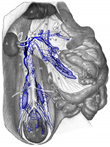 Figure 24: The animal is lying on its back, the abdominal cavity is open, and most of the abdominal organs are laid to the side or pulled out. a liver; b spleen; c pancreas; d jejunum; e ileum; f cecum; g colon; h right kidney; i aorta; k vena cava; l right adrenal gland; m lumbar musculature; n portal vein; o celiac artery; p cranial mesenteric artery; q mesentery with blood vessels; r depressor and s curvator muscles of tail; t deep circumflex iliac artery and vein. 1 right hepatic lymph node; 2 left hepatic lymph node; 3, 31 splenic lymph nodes; 4, 41, 42, 43 jejunal lymph nodes; 5 lumbar aortic lymph nodes; 6 right cranial lumbar aortic lymph node; 7, 71 medial iliac lymph nodes; 8 hypogastric lymph nodes; 9 medial sacral lymph node; 10 lateral sacral lymph node; 11 lumbar trunk; 12 cisterna chyli; 13, 13, 13 lymphatic trunks from the viscera. Source: Dr. Hermann Baum (1918). (This work is in the public domain).
