
Figures 2-7: Schemata of the Left Tracheal Duct
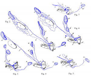
Figures 2-7: a medial retropharyngeal lymph node; b, b’ cranial cervical lymph nodes; c caudal cervical lymph node; d, d’ superficial cervical lymph nodes; e, e’ axillary lymph nodes; f left tracheal duct; g efferent vessels or efferent vessel from the superficial cervical lymph node; i, i’, i”, i'” thoracic duct with its end branches; k lymph vessel from the thyroid 1, that opens into the left tracheal duct f. 1 thyroid gland; 2 axillary vein; 3 external jugular vein; 4 internal jugular vein; 5 1st rib. Source: Dr. Hermann Baum (1918). (This work is in the public domain).
