Sternal Lymph Nodes
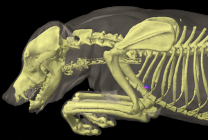
The sternal lymph nodes are shown in Figures 17: c, 18: 6, and 19: 2.
As previously mentioned, the ventral thoracic lymph centre usually consists of a single sternal lymph node on each side. This lymph node can be unpaired and then is found either on one side or in a median location. Rarely, the lymph node is either absent, or 2 are found on one side. In 35 examined cases, there was 1 lymph node on each side in 21 cases, 1 on the right side in 4 cases, 1 on the left side in 3 cases, an unpaired median lymph node in 3 cases, 2 laying above one another on the left side in 2 cases and on the right side in 1 case, and in 1 case absent entirely.
When a lymph node is present on either side, it is found just cranial to the M. transversus thoracis, immediately medial to either the 2nd costal cartilage or the 2nd inter-cartilage space, enclosed in fat, and on the cranioventral border of the internal mammary artery and vein on the sternum (or between the artery and the sternum), filling the space between them with its associated fat. When a single lymph node is present, it may be on either side in the location described above, or it may be located in the middle, ventromedial to the bilateral internal mammary artery and vein, and on a transverse plane at the level of the 2nd costal cartilage or the 2nd inter-cartilage space.
If there are 2 lymph nodes stacked one behind the other on one side, they are both in the location described above, but the caudal lymph node may additionally lie between the internal mammary artery and vein.
The size of the lymph nodes varies between 3 and 20 mm in length.
The absolute weight of all lymph nodes, i.e. the lymph nodes on both sides, ranged between 0.02 and 1.82 g, and the relative weight between 0.005% and 0.009%.
The shape of the lymph nodes is generally oval and somewhat flattened, and the median unpaired lymph nodes are more frequently lobed or dumbbell-shaped. The lymph nodes on both sides can lie so close together that they appear to be a single lymph node.
Afferent drainage
The sternal lymph node drains lymph vessels from the Mm. pectorales, M. serratus ventralis, Mm. intercostales, M. transversus costarum and thoracis, and all the abdominal muscles, the lymph vessels of the ribs and the sternum, the lymph vessels of the diaphragm and mediastinum, the pleura and the peritoneum, the thymus, and the mammary gland. In 1 case in which no sternal lymph node was found, the afferent vessels (at least those that were able to be injected and thus have their drainage pattern assessed), opened into a cranial mediastinal lymph node.
Efferent drainage
One to 3 lymph vessels emerge from each sternal lymph node, forming networks near the internal mammary artery and vein, usually immediately cranioventral to the vessels, and drain to a cranial mediastinal lymph node (almost always the main cranial mediastinal lymph node). If there are 2 lymph nodes located one behind the other on one side, the efferent vessels of the caudal lymph node usually drain partly to the cranial lymph node, with the remainder merging with the efferent vessels of the cranial lymph node.
Clinical Notes