
Figure 20: Lymph Vessels of the Lungs and Tracheobronchial Lymph Nodes of the Dog
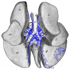
Figure 20: Dorsal side of the lungs; slightly pulled apart. a, a1, a2 left apex, cardiac and base lobes (now known as left cranial [divided into a cranial and caudal part] and caudal lobes); b, b1, b2 right apex, cardiac and base lobes (now known as right cranial, middle and caudal lobes); c Lobus intermedius (accessory lobe); d end of trachea; e left and e’ right main bronchus; f, f pulmonary artery and its branches; g, g pulmonary vein. 1 left tracheobronchial lymph node; 2 right tracheobronchial lymph node; 3 middle tracheobronchial lymph node; 4, 4′ pulmonary lymph nodes; 5 subserous lymph vessel passing around the Margo acutus onto the diaphragmatic surface where it runs deeper; 6, 61 subserous lymph vessels running in the first part of the pulmonary ligament; 7, 7, 7 subserous lymph vessels, which start to run deep at this location; 8 left and 81 right mediastinal lymph node. Source: Dr. Hermann Baum (1918). (This work is in the public domain).
