
Figure 26: Lymph Vessels and Lymph Nodes from the Stomach, Spleen, Pancreas, Duodenum, and Large Intestine of the Dog
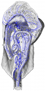
Figure 26: The animal is lying on its back, and the small intestine is removed except for the first part of the duodenum and the end of the ileum. a duodenal lymph node; b right hepatic lymph node; c left hepatic lymph node; d, d’ splenic lymph nodes (a part of the pancreas is cut out so that [c] and [e] became visible); e right colic lymph node; f, f middle colic lymph nodes; g, g left colic lymph nodes; h, h lumbar aortic lymph nodes; i medial iliac lymph nodes (a part of the mesocolon is cut out so that the groups [h] and [i] became visible); k hypogastric lymph node; l lymph vessel of the duodenum and l’ lymph vessel of the pancreas running to the jejunal lymph nodes and therefore cut off; m lymph vessels of the anus and rectum; n lymph vessel running directly to the cisterna chyli; o gastric lymph node; p, p lymph vessels of the rectum passing over the dorsal side of the rectum to the hypogastric and medial iliac lymph nodes. 1 stomach; 2 duodenum (cut off); 3, 3′ pancreas; 4 spleen (with splenic veins laid aside); 5 ileum (cut off); 6 cecum; 7, 8, and 9 colon; 10 rectum; 11 left colic vein; 12 middle colic vein; 13 ileocolic vein (V. ileocaecocolica); 14, 14′ portal vein; 15, 15 ventral wall of the omental bursa, folded back; 16 mesentery of the colon. Source: Dr. Hermann Baum (1918). (This work is in the public domain).
