
Figure 16: Medial Retropharyngeal Lymph Nodes and Cervical Lymph Nodes of the Dog
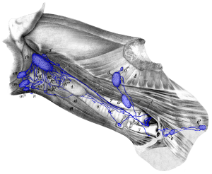
Figure 16: a, a’ medial retropharyngeal lymph nodes; b cranial cervical lymph node; c, c’ caudal cervical lymph nodes; d, d’, d” superficial cervical lymph nodes; e axillary lymph node; e’ accessory axillary lymph node; f left tracheal duct; g efferent vessel of superficial cervical lymph nodes; i thoracic duct with its terminal branches; k, k’, k”, k”‘ lymph vessels from the larynx; l lymph vessel opening into a cranial mediastinal lymph node; m, m1, m2, m3 mandibular lymph nodes; n efferent vessels of mandibular lymph nodes draining to medial retropharyngeal lymph node(s) of the other side. 1 thyroid gland; 2 axillary vein; 3 external jugular vein; 4 internal jugular vein; 5 1st rib; 6 trachea; 7 esophagus; 8 M. serratus ventralis; 9 M. scalenus; 10 M. sternothyroideus; 11 M. sternohyoideus; 12 pharyngeal muscles; 13 M. longus capitis; 14 M. digastricus. Source: Dr. Hermann Baum (1918). (This work is in the public domain).
