
Figure 19: Lymph Vessels of the Pleura of the Dog
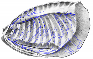 Figure 19: Left thoracic wall and organs of the thorax removed. a, a’ dorsal running lymph vessels; they merge to form larger vessels, which run partly (b) cranial to the cranial mediastinal lymph nodes (1, 1′) and partly (c) caudal to the cranial lumbar aortic lymph node. d, d’ ventral running lymph vessels, which open into the sternal lymph node (2). Some of them (d’) first run over the diaphragm. 1, 1′ cranial mediastinal lymph nodes; 2 sternal lymph node; 3 esophagus (cut off); 4 trachea (cut off); 5 diaphragm (cut off and retracted); 6 M. longus colli; 7 M. transversus thoracis (cut off); 8 left and 8′ right 1st rib; 9 9th rib; 10 13th rib; 11 intercostal lymph node; 12 internal mammary artery and vein. Source: Dr. Hermann Baum (1918). (This work is in the public domain).
