
Lymphosomes of the Dog
Images used with permission from Dr. Hiroo Suami.
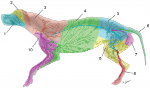
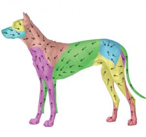
Colour-Coded Diagram of 10 Superficial Lymphatic Territories (Lymphosomes) of the Integument showing Afferent Lymph Vessels and Lymph Nodes (top image), and Direction of Lymph Flow (bottom image): 1 mandibular; 2 parotid; 3 dorsal superficial cervical; 4 axillary; 5 medial iliac; 6 lateral sacral; 7 hypogastric (internal iliac); 8 popliteal; 9 superficial inguinal; 10 ventral superficial cervical. The number of lymph nodes in each lymphosome varied from 1 to 3. The lymph vessels were interconnected within the same lymphosome but not with the neighbouring territory. Source: Suami H, Yamashita S, Soto-Miranda MA, Chang DW. (2013). Lymphatic Territories (Lymphosomes) in a Canine: An Animal Model for Investigation of Postoperative Lymphatic Alterations. PLoS ONE 8(7): e69222 (images used under CC BY 4.0).
