
Figure 17: Lymph Vessels of the Mediastinum, Pericardium, Diaphragm, Aorta, and Esophagus in the Dog
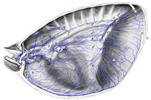 Figure 17: The entire left lung and almost the entire left thoracic wall with the M. transversus thoracis removed. 1, 1 1st rib from which a piece is excised; 2 M. longus colli; 3, 31, 32 aorta; 4 precardiac mediastinum; 5 pericardium and cardiac mediastinum; 6, 61, 62 postcardiac mediastinum; 7, 71, 72 diaphragm; 8 esophagus; 9 cranial vena cava; 10 brachiocephalic artery; 11 left subclavian artery; 12, 12 12th rib from which a piece is excised; 13 13th rib; 14 costocervical vein; 15 thoracic duct; 16 division point of subclavian vein into axillary vein and jugular vein. a, a1, a2 cranial mediastinal lymph nodes; b left tracheobronchial lymph node; b’ middle tracheobronchial lymph node; c sternal lymph node; d, d1 lymph vessels that run to the cranial lumbar aortic lymph node; e lymph vessels that enter the abdominal cavity through the diaphragm and open into splenic lymph nodes, gastric lymph node, left hepatic lymph node or the cranial lumbar aortic lymph node; f lymph vessel entering the abdominal cavity with the esophagus; g intercostal lymph node; h one efferent vessel running to the right side and appearing in Figure 18 (10). Source: Dr. Hermann Baum (1918). (This work is in the public domain).
