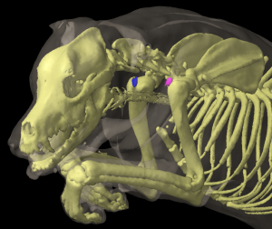
Superficial Cervical Lymph Centre
There is a group of superficial cervical lymph nodes (Figure 13: 3, 3′) on each side of the body, consisting of 2 individual lymph nodes in the majority of cases (16 of 27 dogs examined), though either 1, 3, or 4 lymph nodes may be observed (see below). The lymph nodes are connected to one another and to their surrounding area by loose and usually extremely fatty connective tissue. They lie one above the other, just cranial to the M. supraspinatus on the side of the neck. The lymph nodes are quite superficial and are only covered by the outer skin, the superficial fascia (including the M. cutaneus colli Figure 13: c), and the superficial neck muscles, including the M. trapezius (Figure 13: d), M. brachiocephalicus (Figure 13: g), and M. omotransversarius (Figure 13: e). Medially (Figure 16: d, d’, d”), the lymph nodes abut the M. serratus ventralis (Figure 16: 8), the M. scalenus (Figure 16: 9) and M. longus capitis (Figure 16: 13), and usually the trachea and the esophagus (on the left side). The caudal margin and occasionally the medial surface of the lymph nodes border on the omocervical trunk and the superficial cervical vein and artery. Thin branches from these vessels extend over the lymph nodes, separated from them by fatty connective tissue.
The dorsal lymph node (Figure 13: 3) is usually located just below the skin and the neck fascia, in a triangle formed by the M. trapezius cervicalis (Figure 13: d), M. brachiocephalicus (Figure 13: g) and M. omotransversarius (Figure 13: e). Only the ventral aspect of the lymph node is covered by the M. omotransversarius, while the dorsal end usually extends slightly below the M. trapezius (Figure 13: d).
The ventral lymph node (Figure 13: 3′) is covered by the M. brachiocephalicus (Figure 13: g) and the M. omotransversarius. The ventral lymph node often extends so far ventrally (Figure 16: d’) that the lymph node, or at least its ventral aspect, is medial to the trachea and, on the left side, the esophagus (Figure 16: 7), common carotid artery, internal jugular vein, vagus nerve, and sympathetic nerves, including the depressor nerve and the recurrent nerve.
Exceptions to the number of lymph nodes noted in the 27 examined cases include 1 case in which only 1 lymph node was present on each side (Figure 2: d) and 1 case in which 3 lymph nodes were present on each side (Figure 16: d, d1, d2). Relatively frequently, the number of individual lymph nodes on the left and right side were different; in 2 cases there were 2 lymph nodes on the right and 1 on the left, in 1 case there was 1 on the right and 2 on the left, in 1 case there were 3 on the right and 2 on the left, in 3 cases there were 2 on the right and 3 on the left, and in 2 cases there were 4 on the right (Figure 8: d, d1, d2, d3) and 3 on the left. If 3 lymph nodes are present, they are either stacked on top of one another (as in Figure 16: d, d1, d2), or appear as 2 distinct dorsal lymph nodes and 1 ventral lymph node (Figure 9: d, d1, d2). In the 1 case in which 4 lymph nodes were present on the right side, there was 1 dorsal, 1 ventral and 2 middle lymph nodes (located one behind the other) (Figure 8: d, d1, d2, d3).
The superficial cervical lymph nodes can be quite large. I found lymph nodes in large dogs that were 7.4 cm long, 3.4 cm wide, and 2.1 cm thick. Their shape is also different (as shown in Figures 2-4, 8-10, 11 and 16: d, d1, d2); most of the lymph nodes are oval and somewhat flattened.
The absolute weight of the superficial cervical lymph nodes on both sides varied between 0.32 and 83.92 g, the relative weight between 0.0044% and 0.1583%.
Afferent drainage
The superficial cervical lymph nodes receive afferent lymph vessels from: the skin of the caudal part of the dorsal head region, the pinna, the parotid and neck areas, the caudal half of the cranial neck region, the forelimb digits, the metacarpus, carpus and forearm, most of the lateral side of the upper shoulder, upper foreleg region, and the medial side of the humerus region, the anterior thorax, and the cranial part of the ventral thorax. The lymph nodes also often drain the skin of the masseter region, the antebrachial fascia, most of the shoulder muscles (M. deltoideus, supraspinatus, infraspinatus, teres minor, subscapularis) as well as the Mm. interossei of the forepaw, the extensors and flexors of the 1st and 5th digits, and the tendons of the M. extensor carpi radialis, extensor digitorum communis and lateralis, extensor carpi ulnaris, abductor pollicis longus, as well as the M. flexor digitalis sublimis and profundus, most of the trunk limb muscles (M. trapezius, omotransversarius, brachiocephalic, sternomastoid, rhomboid, serratus ventralis and the Mm. pectorales), the M. splenius, sternohyoideus and sternothyroideus and the neck skin muscle, all bones of the forelimb, the carpal joint, the forepaw digit joints and the pinna.
If 2 to 3 lymph nodes are present, the lymph vessels from distal to mid-brachium (from the skin, muscles, tendons, fasciae, joints and bones) and the shoulder muscles, usually only drain into the ventral superficial cervical lymph node. The lymph vessels from the neck drain into the dorsal (or both dorsal, if multiple are present) lymph nodes, and the lymph vessels from the ventral neck, the skin of the shoulder, and the brachium region drain into both dorsal and ventral lymph nodes.
Efferent drainage
If there is only 1 superficial cervical lymph node (e.g. Figure 2), then 6 to 8 efferent vessels (Figure 2: g) arise from the lymph node, quickly merging to form 1 to 3 vessels that descend over the M. serratus ventralis and scalenus supracostalis. On the left side, the efferent vessels either open into the end of the left tracheal duct (Figure 3: g), into the (frequently bifurcated) end of the thoracic duct (Figure 5: g), into both the left tracheal duct and the thoracic duct (Figure 2, Figure 6: g), or adjacent to the thoracic duct directly into the venous system (common jugular vein) (Figure 7: g). On the right side, the efferent vessels either open into the right tracheal duct (Figure 8: g), unite with the right tracheal duct to form a short right lymphatic trunk (Figure 10, Figure 11: h), or, rarely, open directly into the venous system.
The drainage patterns of the efferent vessels of the superficial cervical lymph nodes have so many variations that it is almost impossible to describe all of them, though I have attempted to illustrate some of the most common patterns in Figures 2-7, 8-12 and in Figure 16: d, d’, d”, and g. If there are several lymph nodes, the 2 to 4 efferent vessels of the dorsal lymph node either enter directly into the ventral lymph node (Figure 9: d2), or combine to form 1 to 2 vessels, which in turn merge with the efferent vessels of the ventral lymph node, forming 1 to 3 small vessels which show the behaviour described above (Figure 3, Figure 4: d, d’, g). I also found cases in which some of the efferent vessels of the dorsal lymph node drained into the ventral lymph node while others merged with the efferent vessels of the ventral lymph node (Figure 6: d, d’), and cases in which the efferent vessels of the dorsal lymph node merged into 1 to 2 vessels which flowed directly to the tracheal duct, or towards the thoracic duct and the common jugular vein (Figure 10: d, d’). Some of these drainage patterns are demonstrated in Figures 2-7, 8-11, and in Figure 16: g.
Clinical Notes
The superficial cervical lymph nodes are commonly called the ‘prescapular’ lymph nodes in the clinic, based on their location cranial to the scapula. Other lymph node groups with common names based on location include the mandibular lymph nodes (‘submandibular’; ventral to the mandible) and the medial iliac lymph nodes (‘sublumbar’; ventral to the lumbar vertebrae).