
Mandibular Lymph Centre
A. Mandibular Lymph Nodes
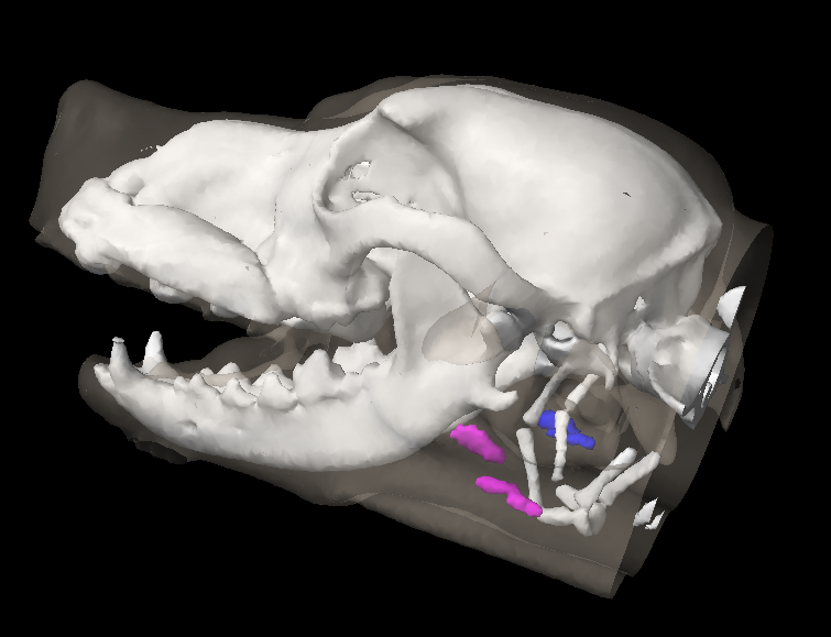
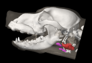
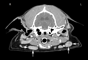
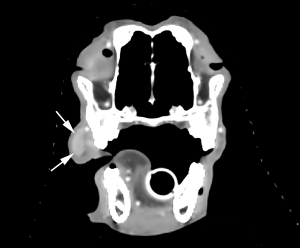
The mandibular lymph nodes (Lnn. mandibulares) (Figure 13: 2, 2′, 2″; 15: v, v1, v2, v3) were examined in 36 dogs. They form a cluster of 2 to 5 lymph nodes on each side. The length of the individual lymph nodes varies from 1.0 to 5.5 cm, the width from 0.75 to 3.0 cm, and the thickness from 0.5 to 1.0 cm. The mandibular lymph nodes are located caudolaterally to the angular process of the mandible, and are underneath the skin, the facial skin muscle, and the superficial fascia.
The external maxillary vein divides the individual lymph nodes into a dorsal and ventral group (Lnn. mandibulares dorsales and ventrales). The dorsal group (Figure 13: 2) may consist of 1 to 2 lymph nodes. If there are 2 lymph nodes present, they rest between the border of the M. masseter and the ramus of the mandible on one side and the mandibular gland on the other, extending to the caudoventral part of the M. masseter and the oroventral border of the mandibular gland. The ventral group (Figure 13: 2′, 2″) consists of 1 to 2 lymph nodes, or rarely of 3, or even 4 lymph nodes, which are located on the M. digastricus, with their ventromedial margin on the M. mylohyoideus and their caudal and ventromedial borders adjoining the lingual vein. As a result, the ventral group of mandibular lymph nodes inserts into the triangle formed by the external maxillary vein and the lingual vein prior to their convergence. In one observed case, the more caudal of the 2 ventral lymph nodes was on the caudoventral border of the lingual vein, i.e. outside the aforementioned triangle formed by the veins, on the lateral surface of the horns of the hyoid bone, or the M. keratopharyngeus.
The number and grouping of the mandibular lymph nodes have been found to be inconsistent, both between different individuals as well as in the same dog between the right and left sides. The numbers of lymph nodes on the right side were as follows: 2 lymph nodes in 19 cases, 3 lymph nodes in 11 cases, 4 lymph nodes in 4 cases, and 5 lymph nodes in 2 cases. On the left side, 2 lymph nodes were found in 17 cases, 3 in 12 cases, 4 in 5 cases, and 5 in 2 cases. In most cases there are 2 lymph nodes on both the right and on the left sides, however, I only found the same number of lymph nodes on both sides in the same individual in 20 cases.
The right dorsal group consisted of 1 lymph node in 33 cases and 2 lymph nodes in 3 cases, and the left dorsal group of 1 lymph node in 31 cases and 2 lymph nodes in 5 cases. The right ventral group was composed of 1 lymph node in 22 cases, 2 in 9 cases, 3 in 4 cases, and 4 in 1 case, while in the left ventral group there was 1 lymph node in 20 cases, 2 in 11 cases, and 3 in 5 cases. I found that in general, there are 1 to 2 mandibular lymph nodes in the dorsal group, where 1 lymph node was found in the majority of cases, present dorsal to the external maxillary vein, and 1 to 3 lymph nodes in the ventral group, where 1 lymph node was found again in the majority of cases, located ventral to the vein. The same number of lymph nodes in both dorsal and ventral groups on the right and left sides were found in 16 cases. In 12 cases, the dorsal and ventral group each consisted of 1 lymph node, in 3 cases the dorsal group consisted of 1 lymph node and the ventral group of 2 lymph nodes, and in 1 case the dorsal and ventral group each consisted of 2 lymph nodes. I therefore cannot agree with Schweitzer [26], who stated that there are always 3 individual lymph nodes in each group.
The absolute weight of the mandibular lymph nodes on both sides varied between 0.2100 and 21.08 g, the relative weight between 0.0082% and 0.044%.
Afferent drainage
The mandibular lymph nodes receive afferent lymph vessels from the skin in the following areas: outer nose, lips, bridge and lateral region of the nose, cheek, intermandibular region, M. masseter, forehead, zygomatic arch, eyelid, parotid region and cranial half (to third) of the front neck region. The lymph nodes also receive lymph vessels from the upper and lower lip, the tip of the tongue, the gums, the cheek, the hard and soft palate, the mucous membrane of the free floor of the oral cavity and the sublingual gland, the outer nose and nasal cavity, the eyelids, lacrimal caruncle, lacrimal gland, zygomatic gland and temporomandibular joint, as well as from various head bones (incisive, nasal, maxillary, frontal, zygomatic, palatine and mandible), various head muscles (M. levator nasolabialis, caninus, levator labii superiorus, zygomaticus, depressor labii inferioris, masseter, temporalis and digastricus, cheek muscles, and M. mylohyoideus), and muscles of the skin of the face and neck.
Efferent drainage
The efferent vessels of the mandibular lymph nodes (Figure 16: m, m1, m2, m3) open into the medial retropharyngeal lymph node (Figure 16: a) and the lateral retropharyngeal lymph node, if present.
However, some of the efferent vessels from the mandibular lymph nodes usually drain to the retropharyngeal lymph nodes on the opposite side (Figure 16: n). Efferent vessels connect the ventral mandibular lymph nodes with each other, and with the dorsal mandibular lymph nodes. I found that the efferent vessels connecting the ventral and dorsal groups only flowed from the dorsal to the ventral group (Figure 13: 2, 2′, 2″; 15: v, v1, v2, v3; 16: m, m1, m2, m3). From each individual lymph node, a large number of efferent vessels arise (usually from 8 to 10), running along the pharyngeal muscles towards the medial retropharyngeal lymph node, forming anastomoses and networks before combining into 3 to 5 vessels and entering the retropharyngeal lymph nodes (Figure 16: m, m1, m2, m3).
A smaller number of efferent vessels leaving the mandibular lymph nodes turn to travel over the surface of the M. sternohyoideus and the pharyngeal muscles and enter the medial retropharyngeal lymph node on the contralateral side. In one observed case, an efferent vessel leaving a dorsal mandibular lymph node ran superficially towards the lateral retropharyngeal lymph node (Figure 15: v, n). According to Schweitzer [26], the mandibular lymph nodes on either side of the head are connected via the efferent vessels, but I could not confirm this finding in any of the numerous cases I examined.
B. Buccal Lymph Nodes
Translator’s note: Baum’s The Lymphatic System of the Dog did not include a description of the buccal lymph nodes (Ln. buccales). These lymph nodes are found in fewer than 10% of dogs, and are located dorsal to the M. zygomaticus and rostral to the M. masseter, in the region of the facial or the superior labial vein.[1]
Clinical Notes
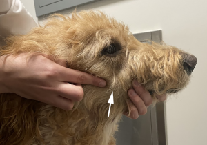
The mandibular lymph nodes are one of three lymph node groups that can often be manually palpated; the other two lymph node groups are the superficial cervical (prescapular) and popliteal lymph nodes. This may allow fine needle aspiration to be performed without imaging guidance.
Some of the efferent vessels of the mandibular lymph nodes always drain to the retropharyngeal lymph node on the opposite side, and therefore the contralateral retropharyngeal lymph node should be assessed in a dog with metastases in a mandibular lymph node.
A normal-sized mandibular lymph node on manual palpation of a dog with oral melanoma should still be cytologically or histologically assessed; in a study of 100 dogs with oral malignant melanoma, 30% of dogs with normal-sized lymph nodes on palpation had evidence of lymph node metastasis on cytology or histology [2]. This finding is likely applicable to other cancers, as early growth of cancer cells within a lymph node may not change the size of the lymph node.
- Casteleyn CR, van der Steen M, Declercq J, Simoens P. The buccal lymph node (lymphonodus buccalis) in dogs: occurrence, anatomical location, histological characteristics and clinical implications. Vet J. 2008 Mar;175(3):379-83. ↵
- Williams LE, Packer RA. Association between lymph node size and metastasis in dogs with oral malignant melanoma: 100 cases (1987-2001). J Am Vet Med Assoc. 2003 May 1;222(9):1234-6 ↵
