
Figure 25: Lymph Vessels of the Small Intestine and the Omentum
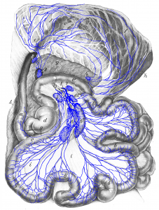 Figure 25: Dog is lying on its back. a duodenum; b, b’ jejunum; c ileum; d cecum; e, e’ colon; f, f omentum (dorsal wall); the stomach shimmers through it; g pancreas; h spleen (largely covered by the omentum); i intestinal mesentery; l cut-open abdominal wall. 1 omental lymph node; 2 duodenal lymph node; 3 right hepatic lymph node; 4 splenic lymph node; 5 right colic lymph node; 6, 61, 62 jejunal lymph nodes; 7 middle colic lymph node; 8 intestinal trunk; 9 lymph vessel of duodenum going into right jejunal lymph node 6; 10 cranial mesenteric artery; 11 jejunal trunk. Source: Dr. Hermann Baum (1918). (This work is in the public domain).
