
Figure 27: Lymph Vessels of the Kidneys and Lymph Nodes Along the Abdominal Aorta and its Terminal Branches
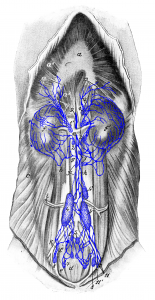
Figure 27: The dog lies on its back, the ventral abdominal wall and the abdominal organs (except for the kidneys) are removed. The kidneys are not drawn in the correct position; the right kidney (f) is pushed back pelvically, so that certain lymph vessels could be drawn into the figure. a, a1, a2 diaphragm; b lumbar musculature; c lateral abdominal wall; d depressor muscles of the tail (muscles that lower the tail) and e lateral flexors of tail; f, f’ kidneys; g caudal vena cava; h abdominal aorta; i right deep circumflex iliac artery and vein; k right external iliac artery and vein; l right hypogastric artery and vein. 1 left and 1′ right cranial lumbar aortic lymph nodes (the latter is covered by the caudal vena cava); 2 lumbar aortic lymph nodes located near the renal artery and vein; 3, 3’ lumbar aortic lymph nodes (those marked [3′] are covered by the caudal vena cava); 4, 41, 42 medial iliac lymph nodes; 5 hypogastric lymph nodes; 6 medial sacral lymph nodes; 7 lateral sacral lymph node; 8, 8′ deep inguinal lymph node; 9 cisterna chyli; 10 pelvic lymphatic trunk; 11 efferent vessels of the superficial inguinal lymph nodes and medial femoral lymph node (some of them [11’] enter the deep inguinal lymph node [8]); 12 lymph vessels which travel with the sympathetic nerve and greater splanchnic nerve from the thoracic cavity into the abdominal cavity; 13 lymph vessels of the diaphragm. Source: Dr. Hermann Baum (1918). (This work is in the public domain).
