Deep Cervical Lymph Centre
The deep cervical lymph nodes (Figures 2, 6, 8, 9, 11, 12 and 16: b, b’, c, c’) are located on the cervical part of the trachea.
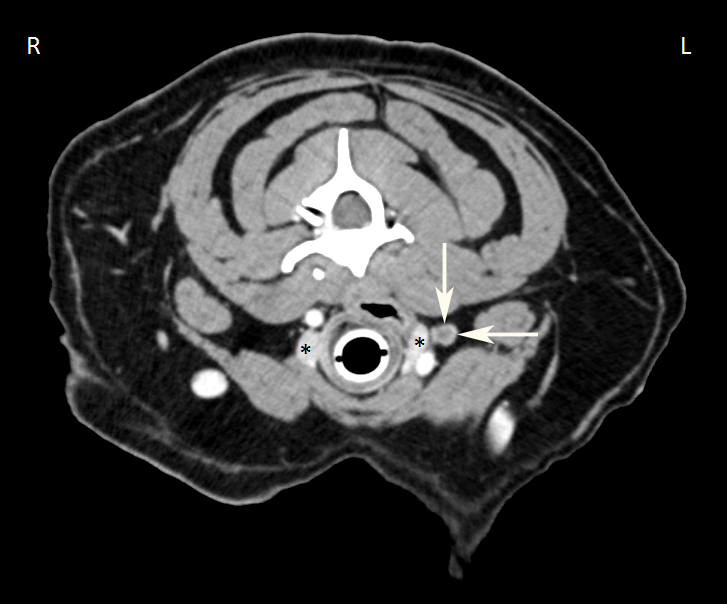
In dogs, in contrast to humans, cattle and horses, the deep cervical lymph nodes are present in sparse numbers, and rarely (3 out of 61 examined dogs) (Figures 4 and 10), absent entirely. If they are present, they can be found along the length of the cervical portion of the trachea. Nevertheless, the deep cervical lymph nodes may be divided into 3 subgroups: the cranial cervical lymph node (Figures 2, 6, 8, 9 and 16: b, b”), the middle cervical lymph node (Figures 8, 12 and 16: c’), and the caudal cervical lymph node (Figures 6, 8, and 16: c). These 3 subgroups may not be present in all cases or may not be clearly demarcated if present. Some of the deep cervical lymph nodes are very small, hardly the size of a pinhead, and can easily be missed on macroscopic examination if the efferent vasculature has not been injected. A range of sizes up to 10 mm was observed, and the shapes are the usual round or oval.
A. Cranial Cervical Lymph Nodes
The cranial cervical lymph node (Figures 2, 6, 8, 9 and 16: b, b’) is located near the thyroid gland (Figure 16: 1), and is found in only approximately one-third of dogs, almost always as a single lymph node. If a lymph node is located near the thyroid gland, between the caudal end of the medial retropharyngeal lymph node and the caudal end of the thyroid gland, I refer to it as a cranial cervical lymph node. More specifically, the lymph node is either on the dorsomedial border of the thyroid gland adjacent to the trachea and common carotid artery (Figure 2: b), or on the craniodorsal aspect of the thyroid gland, between the thyroid gland and the medial retropharyngeal lymph node (Figure 16: b). In the latter case, the cranial cervical lymph node can be found proximate to the pharynx and may even reach the medial retropharyngeal lymph node or overlap it; in these cases, it can be difficult to determine whether the lymph node is a medial retropharyngeal lymph node or a cranial cervical lymph node. I would determine a lymph node in this location to be a cranial cervical lymph node because I have observed in many cases that lymph vessels draining to the medial retropharyngeal lymph node when the cranial cervical lymph node is absent do not drain into the cranial cervical lymph node when it is present. At times, a cranial cervical lymph node lying at the caudal end of the thyroid gland (Figure 11: b) may not be clearly demarcated from the middle cervical lymph node, if present.
The cranial cervical lymph node is absent in most dogs (Figures 4 and 10). It was not found in 45 of 64 cases examined. In the 19 cases in which it was found, there was 1 lymph node on each side (Figures 2 and 16: b) in 12 cases, 1 lymph node on the left in 1 case, 1 lymph node on the right in 3 cases, 1 lymph node on the left and 2 lymph nodes on the right in 1 case (Figure 9: b, b’), and 2 lymph nodes on both sides in 2 cases (Figures 6, 8 and 11: b, b’).
The accessory thyroid glands or the external parathyroid glands may be confused with the cranial cervical lymph node. Microscopic examination will confirm identification, but as the accessory thyroid glands and parathyroid glands lack efferent vessels, injection into the structure to see if it has efferent vessels will determine if it is indeed a lymph node.
Afferent drainage
The cranial cervical lymph node receives afferent lymph vessels from the larynx, thyroid, trachea, and esophagus, and drains the efferent vessels of the medial retropharyngeal lymph node.
Efferent drainage
One to 2 efferent vessels arise from the lymph node, and merge with the left or right tracheal duct (see tracheal duct and Figures 2, 16: b). If there are 2 cranial cervical lymph nodes present, then the efferent vessels of the more caudal lymph node merge with the tracheal duct (Figures 6, 8, 9, 11), while the efferent vessels of the cranial lymph node either open into the caudal lymph node (Figure 6: b), drain into the tracheal duct (Figure 8: b), or show both of the aforementioned behaviours (Figure 9: b).
B. Middle Cervical Lymph Nodes
A middle cervical lymph node (Figures 8, 12, 16: c’; 23: 3), i.e. a lymph node located caudal to the thyroid gland in the central part of the neck near the trachea, was only found in 4 of 50 cases; in 1 case on both sides, in 1 case on the left, in 1 case on the right and 1 case on the ventral aspect of the trachea. In 2 cases, the lymph node was very small, no more than 2 to 3 mm in size, whereas in the other 2 cases, it was 3 to 5 mm in size. These small lymph nodes can be easily missed if the afferent vessels are not injected (see tracheal duct).
Afferent drainage
The middle cervical lymph node receives afferent lymph vessels from the thyroid, trachea and esophagus.
Efferent drainage
On average, 1 efferent vessel arises from the lymph node, coursing caudally alongside the trachea and draining into either the caudal cervical lymph node (Figures 8, 16: c’, c; 23: 3, 4), the end of the thoracic duct, the right lymphatic duct, the tracheal duct (Figure 12: c’), or into a cranial mediastinal lymph node.
C. Caudal Cervical Lymph Nodes
The caudal cervical lymph node (Figures 6, 8, 16: c; 23: 4) is also absent in the majority of cases, and in 39 of 56 examined cases, it could not be detected at all. In the 17 cases in which the lymph node was present, it was located at the caudal end of the cervical trachea within 4 cm cranial of the neck of the 1st rib, covered by the anterior neck muscles (M. sternohyoideus, sternothyroideus and brachicephalicus). Of the 17 cases with a lymph node present, there was 1 unpaired lymph node on the ventral side of the trachea (11 cases), 1 lymph node on each side of the trachea (2 cases), 1 lymph node on the left side and 1 lymph node on the ventral side of the trachea (1 case), or either 3 (2 cases) or 4 (1 case) consecutive lymph nodes on the ventral side of the trachea. In the case of 4 lymph nodes, all of which were only 1.5 to 2.5 mm in size, the cranial lymph node may instead have been a middle cervical lymph node, as the demarcation between the two subgroups is minimal. In another case, the lymph node found was only the size of a pinhead – so small that it could easily have been overlooked (see cranial cervical lymph nodes, above).
Afferent drainage
If the caudal cervical lymph node(s) are present, some of the lymph vessels that would typically enter the thoracic cavity and drain to the mediastinal lymph node instead drain into the caudal cervical lymph node(s). These include lymph vessels draining muscles such as the M. splenius, sternohyoideus, sternothyroideus, sternocostalis, scalenus, serratus ventralis, longus colli, longus capitis, and more. However, not all the lymph vessels draining these muscles enter the caudal cervical lymph node(s); some bypass it and drain into the mediastinal lymph node. Direct drainage into the caudal cervical lymph node(s) was observed for the last 5 to 6 cervical vertebrae, the thyroid, trachea and esophagus, and the efferent vessels of the middle cervical lymph node(s).
Efferent drainage
Each caudal cervical lymph node has 1 to 2 efferent vessels. If there is more than 1 lymph node, the efferent vessel of the most cranial lymph node usually drains into the lymph node caudal to it, and only the efferent vessel of the most caudal lymph node (or if there is only one lymph node, the efferent vessel from it), opens either into the end of the thoracic duct (Figure 6: c), into the right lymphatic trunk (on the right side) (Figure 8: e), into the end of the tracheal duct, or into a cranial mediastinal lymph node (Figures 16: c, l; 23: 4, 6). In the latter case, the efferent vessels often form extensive networks.
Clinical Notes