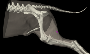
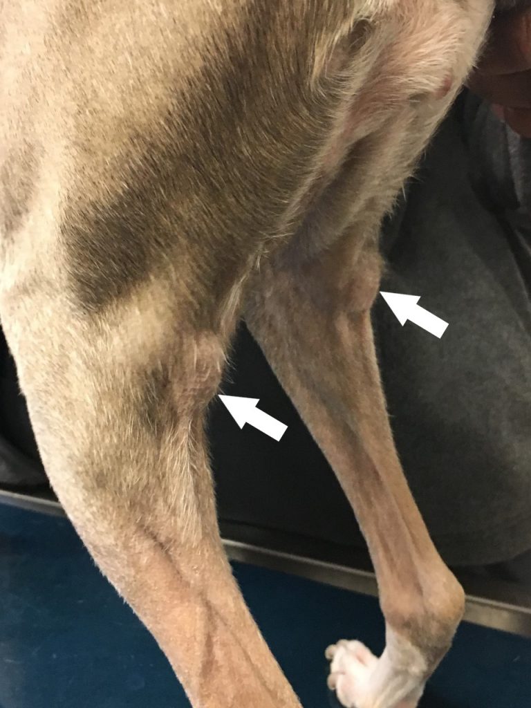
Popliteal Lymph Nodes
The popliteal lymph node (Figure 13: 5) is almost always a single lymph node, as 2 popliteal lymph nodes on the right side were found in only 1 of 37 cases. In large dogs, the popliteal lymph node can reach a length of 5.0 cm, a width of 3.4 cm, and a thickness of 1.4 cm.
The location of the lymph node is just as constant as its quantity. It lies in the caudal aspect of the stifle joint between the M. biceps femoris and M. semitendinosus (Figure 13: p, q), on the caudal surface of the M. gastrocnemius (Figure 16), and is usually within an ample amount of fatty tissue. Since the lymph node usually protrudes slightly caudally over the M. biceps femoris and M. semitendinosus (shown in Figure 13), the lymph node, together with the fat pad between the two muscles, comes into direct contact with the skin.
The absolute weight of the lymph node on both sides varied between 0.04000 and 19.07 g, the relative weight between 0.0007% and 0.036%.
Afferent drainage
The popliteal lymph node drains lymph vessels from the skin of the caudal half of the lateral side of the stifle joint and lower leg, the lateral, flexor, and extensor aspects of the tarsus, the skin of the metatarsus and the hind digits, the lymph vessels from the fascia cruris, the tibia, the fibula, the tarsal and metatarsal bones, the phalanges of the toes, the tarsal joint and the interphalangeal joints, the lymph vessels from the M. biceps, semitendinosus, semimembranosus, quadriceps, extensor digitalis pedis brevis, and the Mm. interossei, and the lymph vessels from the tendons of the M. tibialis anterior, extensor digitalis pedis longus and lateralis, peroneus longus, gastrocnemius, and flexor digitalis pedis sublimis and profundus.
Efferent drainage
Eight to 10 efferent vessels emerge from the popliteal lymph node (Figure 34: a’) and, accompanied by the small saphenous vein, run along the medial side of the M. biceps and gastrocnemius to the caudal aspect of the stifle joint, forming networks and finally merging to form 2 to 4 vessels. These vessels then travel medially, just above the origin of the M. gastrocnemius, in the angle between the M. semimembranosus, the M. adductor, and the femur, through the femoral canal to the medial iliac lymph node. If either a medial femoral lymph node or deep inguinal lymph node is present, one of the efferent vessels usually enters it.
In one case where a left popliteal lymph node was observed, in addition to the efferent vessels described above, I found another efferent vessel that arose directly under the skin at the border between the M. biceps and semitendinosus, entered the pelvic cavity and drained into the left medial sacral lymph node. This left medial sacral lymph node drained to the hypogastric lymph nodes and then, via the efferent vessels of the hypogastric node, to both medial iliac lymph nodes. In this pattern, lymph from the left popliteal lymph node drained into the left medial sacral lymph node, the hypogastric lymph nodes, and both of the medial iliac lymph nodes.
Clinical Notes