Bronchial Lymph Centre
The bronchial lymph centre includes all lymph nodes located on the bifurcation of the trachea and the bronchi: (1) the tracheobronchial lymph nodes (previously the Lnn. bifurcationis) (Figures 20 and 22: 1, 2, 3), which include the left (Figure 22: 1), right (Figure 22: 1) and middle (Figure 22: 1) tracheobronchial lymph nodes, which lie on the left, right and caudal side of the bifurcation, respectively, and (2) the pulmonary lymph nodes (Figures 20 and 22: 4, 4′), which lie on the bronchi before they enter the lungs, almost always on their dorsal side. I call these pulmonary lymph nodes even though they are not located in the lungs, because they only receive lymph vessels from the lungs, and because they correspond to the pulmonary lymph nodes located on the bronchi in cattle, although in that species the lymph nodes are within the lungs (Baum [6] page 36). I also have chosen not to divide the lymph nodes located on the bifurcation of the trachea and the bronchi into as many separate subgroups as is done in man, in whom the lymph nodes include the bifurcation lymph node (at the angle of the bifurcation), the tracheobronchial lymph nodes (at the angles between the trachea and the bronchi), the bronchopulmonary lymph nodes (at the branching angles of the bronchi), and the pulmonary lymph nodes (in the lung tissue).
Such an extensive subgrouping of the bronchial lymph nodes cannot be applied to the anatomy of other species and would create confusion as to which subgroup each lymph node should be assigned to. The pulmonary lymph nodes in the dog are comparable to the bronchopulmonary lymph nodes in man. The descriptions I provide refer only to the lymph nodes that are visible macroscopically and not to microscopically detectable lymph nodes or the smallest lymph nodes.
A. Tracheobronchial Lymph Nodes
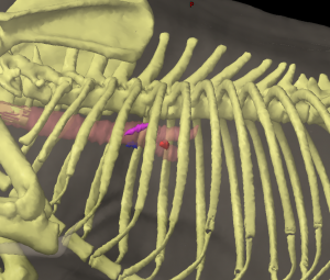
The tracheobronchial lymph nodes (Figures 17: b, b’, 18: 1, 2, 20 and 22: 1, 2, 3) are fairly constant in number and position. In 34 closely examined cases, there were always 3 lymph nodes: 2 of the lymph nodes were consistently found at the right and left bifurcation, lateral to the border between the trachea and bronchi (the right and left tracheobronchial lymph node, respectively), and the 3rd lymph node in bifurcation angle (the middle tracheobronchial lymph node). The size of the lymph nodes ranged from 6 mm to 3.2 cm. The middle tracheobronchial lymph node was always the largest. The lymph nodes are conspicuous because they are mostly black in colour (due to the deposition of dust and other particles through filtration).
The right tracheobronchial lymph node (Figures 18, 20, and 22: 2) lies in the angle between the trachea and the right main bronchus, cranial to the bronchus for the right apical lobe (currently known as the right cranial lobe) of the lung (as shown in Figure 20) and often contacting the bronchus with its caudal surface (Figure 18: s’). The lymph node is bordered craniodorsally by the azygos vein (Figure 18: h), and ventrally by the branches of the pulmonary artery and vein that supply the right apical lung lobe (right cranial lung lobe). The right apical lung lobe (right cranial lung lobe) borders the right tracheobronchial lymph node laterally.
The left tracheobronchial lymph node (Figures 17: b, 20; 22: 1) is in a comparable position to the right lymph node, in the angle between the trachea and the left main bronchus. The left lymph node is bordered craniodorsally by the thoracic aorta (Figure 17: 3), though its cranial part may extend between the trachea and the aorta. Ventrally, it is bordered by the left branch of the pulmonary artery. The lymph node’s left side, which is covered by the left apical lung lobe (currently known as the cranial segment of the left cranial lung lobe), is crossed by the left vagus nerve.
The middle tracheobronchial lymph node (Figures 17: b’; 18: 1; 20, and 22: 3) is located in the bifurcation angle and extends from one main bronchus to the other in a slightly convex, cranial arc, so that it is almost horseshoe-shaped, with a middle section and two legs (see Figure 20: 3). It also extends slightly outwards on the dorsal aspect of the two main bronchi. It is bordered dorsally by the esophagus and caudoventrally by the pulmonary veins, and it is crossed by the vagus nerves laterally on both sides.
The absolute weight of all the tracheobronchial lymph nodes ranged from 0.25 g to 6.78 g, and the relative weight from 0.0011% to 0.0256%.
Afferent drainage
The tracheobronchial lymph nodes drain lymph vessels from the thoracic portion of the esophagus, the end of the trachea, the bronchi and lungs, the mediastinum and diaphragm, the heart, and the aorta, as well as the efferent vessels of the pulmonary lymph nodes.
Efferent drainage
Two to 4 efferent vessels arise from each tracheobronchial lymph node, some of which drain into another tracheobronchial lymph node, and some of which drain into 1 or more mediastinal lymph nodes. Some of the efferent vessels of the right tracheobronchial lymph node travel over the dorsal and ventral surfaces of the right main bronchus into the middle tracheobronchial lymph node, and others drain into the right mediastinal lymph node (Figures 18: u; 20: 2). Some of the efferent vessels of the middle tracheobronchial lymph node (Figures 17: b’; 18: 1 and 20: 3) travel both dorsally and ventrally over the left main bronchus and drain to the left tracheobronchial lymph node, and some drain directly to the mediastinal lymph nodes on both the right and left sides. In one case, an efferent vessel from the middle tracheobronchial lymph node passed over the left tracheobronchial lymph node without opening into it. One to 2 efferent vessels from the left tracheobronchial lymph node (Figures 17: b; 20: 1) travel over the left side, or over both sides, of the aortic arch to one of the left mediastinal lymph nodes, usually either to the lymph node located on the left side of the cranial vena cava (Figure 17: a’), or to the lymph node located on the brachiocephalic artery, sometimes to both aforementioned lymph nodes. In 3 cases, none of the efferent vessels from the left tracheobronchial lymph node drained into either the middle or right tracheobronchial lymph nodes, or at most there was drainage only observed up to the 1st valve. Drainage from the middle tracheobronchial lymph node to the right tracheobronchial lymph node was also rare.
B. Pulmonary Lymph Nodes
The pulmonary lymph nodes (Figures 20, 22: 4, 4’) are usually small, ranging from 4 to 10 mm in length, although, in rare cases, they may be considerably larger (up to 20 mm in length). The lymph nodes are located on the extrapulmonary aspect of the left and right bronchi, usually on the dorsal side. The pulmonary lymph nodes are not consistently found – on the contrary, they are absent in the majority of cases; in 41 examined cases, they were found in only 14. Of these 14 cases, a lymph node on the right bronchus was present in 10, and a lymph node on the left bronchus was present in 4. In 1 of the latter cases, the node seemed to originate from the left side of the middle tracheobronchial lymph node. The pulmonary lymph nodes are usually black in colour due to the deposition of dust and other particles.
Afferent drainage
The pulmonary lymph nodes drain lymph vessels from the bronchi and the lungs.
efferent drainage
One to 2 efferent vessels arise from each pulmonary lymph node and drain into either an ipsilateral tracheobronchial lymph node or mediastinal lymph node, or to both a tracheobronchial and mediastinal lymph node, as shown in Figure 20: 4, 4’.
Clinical Notes