
Figure 36: Lymph Vessels of the Joints of the Forelimb of the Dog (Medial Side)
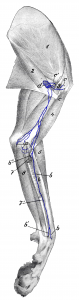
Figure 36: a lymph vessels from the elbow joint; b, b’, b” lymph vessels from the carpal joint (b” emerges from under the M. pronator quadratus); c, c’ lymph vessels of the shoulder joint (the one labeled c’ comes from the lateral side of the shoulder joint); d axillary lymph node. 1 M. subscapularis; 2 M. teres major; 3 M. coracobrachialis; 4 M. biceps; 5 M. extensor carpi radialis; 6 M. pronator quadratus; 7, 7′ M. flexor carpi radialis (a piece is cut out); 8 M. flexor digitalis sublimis. Source: Dr. Hermann Baum (1918). (This work is in the public domain).
