
Figure 29: Lymph Vessels of Reproductive Organs of the Female Dog
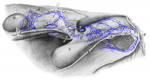 Figure 29: 1 medial iliac lymph node; 2 and 3 lumbar aortic lymph nodes; 4 hypogastric lymph node; 5 superficial inguinal lymph nodes; 7 lateral sacral lymph node; 8 lymph vessel bending over to the other surface of the uterine horn. a left kidney (folded back); b left ovary embedded in fat (folded back); c left uterine horn (folded back); c’ right uterine horn; d uterine body; e vagina; f vaginal vestibule; g vulva; h urinary bladder; i urethra; k suspensory ligament of the ovary (ligamentum suspensorium ovarii) including the ovary-kidney ligament; l broad ligament of uterus (ligamentum latum uteri); m lateral bladder ligament; n ventral abdominal wall (cut lateral to the linea alba); o, o’ ventral pelvic wall (cut left of the median plane). Source: Dr. Hermann Baum (1918). (This work is in the public domain).
