
Figure 32: Lymph Vessels of the Urinary and Reproductive Organs of the Male Dog
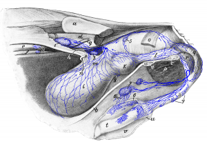 Figure 32: The left abdominal and pelvic wall and the intestine are removed; the penis is slightly detached from the ventral abdominal wall. 1 medial iliac lymph node; 2 lumbar aortic lymph nodes; 3 hypogastric lymph node; 4 superficial inguinal lymph nodes; 5 lateral sacral lymph node; 6, 6′ lymph vessels of the integument of the prepuce; 7 lymph vessels of the urethra; 8 lymph vessel of the urinary bladder, which bends to the other side. a ilium (sawn-off); b ventral abdominal wall (cut left of the linea alba); c, c ventral pelvic wall (cut left of the median plane); d lumbar musculature; e aorta; f vena cava; g left external iliac artery; h left hypogastric artery; i urinary bladder; k prostate; l urethra; m lateral ligament of the urinary bladder; n ureter; o M. coccygeus (cut off); p cut surface from M. adductor; q M. bulbocavernosus; r M. ischiocavernosus; s penis; t glans; u cavernous node; r prepuce (opened and retracted). Source: Dr. Hermann Baum (1918). (This work is in the public domain).
