Medial Iliac Lymph Nodes
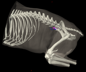
The medial iliac lymph node (Figure 24: 7, 7’; 26: 1; 27: 4, 41, 42; 32: 1) is usually found as one elongated, slightly flattened lymph node on each side of the body. It is adjacent to the aorta on the left side (Figures 24: 7’; 27: 41, 42) and to the caudal vena cava on the right side (Figure 24: 7; 27: 4), between the deep circumflex iliac artery (Figure 27: i) and the external iliac artery. The lymph node is 6 cm long, 2.2 cm wide, and 1.5 cm thick in large dogs. In 13 of 33 cases, the number of lymph nodes deviated from this description: in 2 cases there was 1 lymph node on the left and 2 on the right, in 8 cases 1 on the right and 2 on the left, in 2 cases 1 on the right and 3 on the left, and in 1 case, 2 lymph nodes each on both the right and left sides. Since the number of cases in which there was more than 1 lymph node on each side is quite high, one could speak of the medial iliac lymph node in the plural. I do not, however, because the cases in which there is only 1 lymph node on each side is still predominate in number, and because the lymph vessels from one limb only open into the lymph node on the ipsilateral side, and were not observed to cross the median plane and flow into the contralateral lymph node. The medial iliac lymph node is usually ventral to the bodies of the 5th and 6th lumbar vertebrae and ventromedial to the lumbar muscles at the angle between the aorta and the cranial edge of the external iliac artery on each side.
The right lymph node (Figure 24: 7; 27: 4) lies on the side of the abdominal aorta, ventral to the caudal vena cava and right common iliac vein. This lymph node usually starts just caudally from the deep circumflex iliac artery (Figure 27: i) and extends to the right external iliac artery (Figure 27: k), at the cranial edge of which it sometimes bends laterally to accompany the artery for a short distance. Rarely, part of the lymph node is located to the right of the caudal vena cava on the ventromedial side of the flat tendon of the M. psoas minor. Quite often, the cranial one-quarter or one-third of the lymph node protrudes over the deep circumflex iliac artery, so that it is crossed by the artery on its dorsal side (as Figure 27 shows). The lymph node may also extend so far caudally that it lies between the external iliac artery and the common iliac vein on the right side, and is therefore crossed on its ventral side by the external iliac artery.
The left lymph node (Figure 24: 7’; 27: 41, 42) is usually found at the angle between the abdominal aorta and the cranial edge of the left external iliac artery and frequently extends to some degree along the latter. The lymph node extends dorsally to the tendons of the M. psoas minor and the M. iliopsoas and usually extends from the deep circumflex iliac artery to the left external iliac artery. Not infrequently, however, the cranial one-quarter to one-third of the left lymph node protrudes beyond the deep circumflex iliac artery (as shown in Figure 24), and may extend in a caudal direction over the dorsal side of the external iliac artery to the hypogastric artery, or up to, or even over, the dorsal aspect of the common iliac vein. In cases where 2 or 3 lymph nodes are present on one side, they are generally found together, one behind the other, and were only rarely observed to be positioned next to each other. In these cases, either 1 lymph node is located just cranial to the deep circumflex iliac artery and the other lymph node just caudal to it (as shown in Figure 24), or 1 lymph node extends from the deep circumflex iliac artery to the external iliac artery and the other is located caudal to it.
If a medial iliac lymph node is located just cranial to the deep circumflex iliac artery, it may be difficult to distinguish from a lumbar aortic lymph node, and it is possible that it should be identified as such. In support of this being a lumbar aortic lymph node, it was found that when such a lymph node is present, the lymph vessels ascending in the femoral canal do not drain into it, but drain first into the caudal of the 2 lymph nodes, and only through the efferent vessels of the caudal lymph node does the lymph reach this lymph node. However, in support of the lymph node being a medial iliac lymph node, frequently there is only 1 lymph node that protrudes cranially over the deep circumflex iliac artery in such a way that the cranial portion of the lymph node is only connected to the rest of the lymph node by a parenchymal bridge. Thus, one could get the impression that if the parenchymal bridge were absent, the cranial portion would be considered an independent medial iliac lymph node just cranial to the deep circumflex iliac artery. In 1 case with 3 lymph nodes, 1 lymph node was dorsal to the deep circumflex iliac artery, 1 lymph node was ventral to the deep circumflex iliac artery, and the 3rd lymph node was caudal to the deep circumflex iliac artery on the craniolateral border of the external iliac artery. For the absolute and relative weight of the medial iliac lymph node see the hypogastric lymph nodes.
Afferent drainage (Figures 24, 26, 27, 29, 31, and 32)
The medial iliac lymph node drains lymph vessels from the skin of the dorsal half of the part of the abdominal wall that is located caudally from a transverse plane cutting through the last rib, from the skin of the pelvic region and the base of the tail and the cranial half of the lateral side of the thigh and stifle joint, lymph vessels from the fascia lumbodorsalis, fascia lata and fascia cruris, lymph vessels of all muscles in the pelvis, thigh, lower thigh and foot, including the lumbar muscles with the exception of the M. obturatorius internus, lymph vessels of the tendons of the M. tibialis anterior, M. extensor digitalis lateralis, M. peroneus longus, M. gastrocnemius, M. flexor digitalis sublimis and profundus, lymph vessels of all abdominal muscles and of the abdominal skin muscle, lymph vessels of the hip, knee and tarsal joints, lymph vessels of the pelvis, femur, patella, tibia and fibula and tarsal and metatarsal bones, lymph vessels of the colon, rectum and anus, lymph vessels of the uterus, vagina, vaginal vestibule and vulva, lymph vessels of the testicles, epididymis, ductus deferens, tunica vaginalis communis, M. cremaster, the prostate and the male and female urethra, lymph vessels of the urinary bladder and ureter, lymph vessels of the aorta, the peritoneum and the nervous system, as well as the efferent lymph vessels of the popliteal lymph node, medial femoral lymph node, deep inguinal lymph node, superficial inguinal lymph nodes, left colic lymph nodes, sacral lymph node, and hypogastric lymph nodes.
Efferent drainage (Figures 24 and 27)
The efferent vessels of the medial iliac lymph node merge to form several trunks, some of which drain to a lumbar aortic lymph node, and some of which merge with the efferent vessels of the lumbar aortic lymph nodes to form the pelvic lymphatic trunk. The medial iliac lymph nodes on each side are connected to each other by efferent lymph vessels, as are the lymph nodes on one side if there is more than one present.
Clinical Notes
The medial iliac lymph nodes on each side are connected to each other by efferent lymph vessels, and cancer cells shed from a malignant tumour on one side could therefore travel to the medial iliac lymph node(s) on both sides.