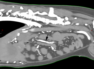
Jejunal Lymph Nodes
The jejunal lymph nodes (Figure 25: 6, 61, 62) are the lymph nodes of the jejunum and ileum. They are fairly constant in number and location. They were frequently found to consist of 2 elongated, flattened nodes that taper to somewhat of a point at both ends and were located between the sheets of the long jejunal mesentery, near the jejunal arterial and venous trunks (Figure 25: 11). Both lymph nodes usually extend from the cranial mesenteric root to the point where the trunks of the jejunal arteries and veins divide into their terminal branches. One of the lymph nodes (Figure 25: 6) is usually dorsal and slightly to the right of the aforementioned vascular trunks and is therefore referred to as the right jejunal lymph node, while the second (Figure 25: 61, 62) is located to the left of the other lymph node and ventrally to the vessels, and is thus referred to as the left jejunal lymph node (see also Figure 24: 4, 41, 42, 43).
The right jejunal lymph node (Figure 25: 6) usually lies between the trunk of the jejunal veins and the ileum and can be seen more clearly from the dorsal side of the mesentery. It usually extends to the right colic lymph node (Figure 25: 5), and there are often 2, or even 3, right jejunal lymph nodes. If there are 2 lymph nodes, the more caudal lymph node is the main lymph node – it may also be discernible as the much larger one. The other lymph node, which is much smaller, is cranial to the main lymph node but is usually still found either between the trunk of the jejunal vein and the ileum, or directly on the trunk of the vein. If there is a 3rd lymph node, it is also quite small and either lies in front of the trunk of the aforementioned vein, or on the border of the vein facing away from the ileum. Occasionally, the smaller lymph nodes are located between the main lymph node and the ileum. In 25 cases examined, 1 lymph node was found in 20 cases, 2 lymph nodes were found in 4 cases, and 3 lymph nodes were found in 1 case.
The left jejunal lymph node (Figure 25: 61) can be seen more clearly from the ventral view of the mesentery. It sometimes extends to the ventral aspect of the middle colic lymph node (Figure 25: 7). Additionally, there may also be 1 to 2 smaller lymph nodes, either cranial to the main left jejunal lymph node (Figure 25: 62) or caudal to it. In 1 case, there were 2 smaller cranial and 2 smaller caudal lymph nodes in addition to the main lymph node, so that there were a total of 5 lymph nodes on the left side. In 25 cases examined, there was 1 lymph node in 21 cases, 2 lymph nodes in 2 cases, 3 lymph nodes in 1 case, and 5 lymph nodes in 1 case.
The sizes of the jejunal lymph nodes range from 0.5 cm to 20 cm in length, 4 mm to 2 cm in width, and 3 mm to 1 cm in thickness.
Afferent drainage
The jejunal lymph nodes drain lymph vessels from the jejunum, ileum, and pancreas.
Efferent drainage
A very large number of efferent vessels emerge from each of the larger jejunal lymph nodes (Figures 24: 4, 41, 42, 43; 25: 6, 61, 62). These vessels merge with the efferent vessels from neighbouring jejunal lymph nodes, as well as with the efferent vessels from the right colic, middle colic, hepatic, and splenic lymph nodes, forming an extensive vascular network. From this network, several larger lymph vessels arise and merge to form the intestinal trunk(s) (see intestinal trunk). Due to this network, when the efferent vessels of one lymph node are injected, the injected material fills the efferent vessels of other lymph nodes. Usually, the efferent vessels of the right jejunal lymph node also drain to the right colic lymph node, and the efferent vessels from the left jejunal lymph node drain to the middle colic lymph node, just as the jejunal lymph nodes are connected with each other.
Clinical Notes


