
Figure 23: Lymph Vessels of the Cervical Part of the Trachea and Esophagus of the Dog
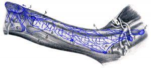 Figure 23: Parts located in the thoracic cavity are shown in Figure 17. a, a cervical part of the trachea; b, b cervical part of the esophagus; c veins (axillary vein, external and internal jugular veins); d M. sternothyroideus; e M. sternohyoideus; f M. thyropharyngeus and cricopharyngeus; g M. hypothyroideus; h M. keratopharyngeus. 1 medial retropharyngeal lymph node; 2 cranial cervical lymph node; 3 middle cervical lymph node; 4 caudal cervical lymph node; 5 lymph vessels from the initial part of the esophagus; 6, 6′ cranial mediastinal lymph nodes; 7, 7 1st rib from which a piece is excised. Source: Dr. Hermann Baum (1918). (This work is in the public domain).
