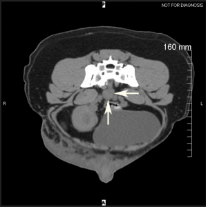
Sacral Lymph Nodes
The sacral lymph nodes (Figures 24: 9, 10; 27: 6, 7; 29: 7; 32: 5) are lymph nodes located on the inner aspect of the roof and dorsolateral walls of the pelvic cavity, caudal to the hypogastric lymph nodes, and are sometimes very difficult to distinguish from the hypogastric nodes. The sacral lymph nodes only occur in about half of all cases and, moreover, are easy to overlook because they are usually small lymph nodes that are always enclosed in fat. The sacral lymph nodes can be divided into the medial sacral lymph node, located at the roof of the pelvic cavity, and the lateral sacral lymph node, located at the dorsolateral wall of the pelvic cavity.
A. Medial Sacral Lymph Nodes
The medial sacral lymph node (Figures 24: 9; 27: 6) lies caudal to the body of the last lumbar vertebra on either the ventral surface of the sacrum or the M. sacrococcygeus ventralis, immediately lateral to the median sacral artery (as shown in Figure 27), which it sometimes covers. The medial sacral lymph node may be located so close to the sacral promontory that it cannot be clearly separated from the hypogastric lymph nodes. In 30 closely examined dogs, medial sacral lymph node(s) were found in 17 cases: 6 times on both sides, 4 times only on the left side, and 2 times only on the right side. In 5 cases, there was only 1 lymph node located on the median, directly on the ventral side of the median sacral artery (as shown in Figure 23). In 2 cases, the lymph node was doubled on one side. The size of the lymph node ranged from 3 to 15 mm. For the absolute and relative weights, see lateral sacral lymph nodes (below).
Afferent drainage (Figures 24, 26, 27, 29, 31, and 32)
The afferent drainage area is difficult to determine due to the inconsistent occurrence of the lymph node. It has been shown that lymph vessels from the tail muscles, sacrum, caudal vertebrae and pelvis (ischium) flow into the lymph node. No lymph vessels of the muscles of the pelvis and thigh drained into the lymph node, although lymph vessels of several muscles (e.g. the M. biceps and M. semitendinosus), accompanied by the sciatic nerve, ran near the lymph node but did not enter it.
Efferent drainage
One to 3 efferent vessels arise from each lymph node, some of which always drain to the hypogastric lymph nodes, some to the medial iliac lymph node, and some rarely to the lateral sacral lymph node (as shown in Figures 26 and 34).
B. Lateral Sacral Lymph Nodes
The lateral sacral lymph node (Figures 24: 10 and 27: 7) lies on each side on the medial side of the M. piriformis in the fat between the M. sacroccygeus ventralis and the M. coccygeus. Usually, the lymph node is partially inserted between the two muscles and is bordered by the hypogastric artery and vein. In 30 dogs examined in more detail, the lateral sacral lymph node was found in only 8 dogs: 6 times on both sides and 1 time each only on the left and right side. In 2 cases, there were 2 lymph nodes lying one behind the other on the left side.
The size of the lymph node ranged from 3 to 14 mm. For the relative and absolute weights, see below.
Afferent drainage
As with the medial sacral lymph node, the infrequent occurrence of the lateral sacral lymph node makes it is very difficult to determine the afferent drainage area. It was shown that the lateral sacral lymph node drains lymph vessels from the sacrum, the caudal vertebrae, the pelvis and femur, from the elevator muscles of the tail and the Mm. gemelli, from the uterus, vagina, vaginal vestibule, vulva and clitoris, the prostate, penis, and the male and female urethra. Of the muscles of the pelvis and thigh, only the lymph vessels of the Mm. gemelli drained to the lateral sacral lymph node, although lymph vessels draining other muscles (e.g. the M. gluteus medius), together with the sciatic nerve, came near the lymph node but did not enter it.
efferent drainage
Some of the efferent lymph vessels of the lateral sacral lymph node drain to the hypogastric lymph nodes, some to the medial iliac lymph node, and sometimes, some also drain to the contralateral medial or lateral sacral lymph node. The vessels also form a complex network caudal to the hypogastric lymph nodes on the ventral side of the last lumbar vertebra (Figures 24 and 27).
The absolute and relative weights were determined for all sacral lymph nodes found on both sides. The absolute weight ranged from 0.01 g to 3.13 g, and the relative weight from 0.0004% to 0.0057%.
Clinical Notes


