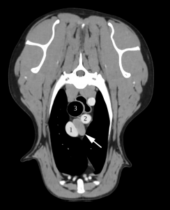
Mediastinal Lymph Nodes
Mediastinal lymph nodes, i.e. lymph nodes located in the mediastinum, occur infrequently in dogs, in much fewer numbers and in much fewer groups than in cattle. While in cattle there are cranial, middle, caudal, dorsal, and ventral mediastinal lymph nodes (see Baum [6] page 29), in dogs there are only the cranial mediastinal lymph nodes, i.e. lymph nodes that lie between the 1st rib and the heart in the precardiac mediastinum.
The cranial mediastinal lymph nodes are sometimes joined by a lymph node, which is, strictly speaking, a middle mediastinal lymph node, as it lies in the cardiac mediastinum. This lymph node is sometimes found on the left side of the pericardium, where the aortic arch emerges, near the cranial border of the pericardium (Figure 17: a2), and sometimes on the right or dorsal side of the trachea, just cranial to the azygos vein (Figure 18: 32), at times partly or entirely between the trachea and azygos vein. The middle mediastinal lymph node only rarely occurs, is usually poorly distinguishable from the cranial mediastinal lymph nodes, and shares the same afferent drainage. For these reasons, the middle and cranial mediastinal lymph nodes are described together in the following sections, and the term mediastinal lymph nodes is used, but these descriptions are almost exclusively of the behaviour of the cranial mediastinal lymph nodes.
One of the mediastinal lymph nodes is notably constant in its occurrence and position and lies in the 1st intercostal space, just cranial to the costocervical vein. This lymph node will be described in more detail below, and to facilitate the description of its afferent drainage, it will be referred to as the main (cranial) mediastinal lymph node. A caudal mediastinal lymph node was not found in any of the numerous cases examined. The tracheobronchial (bifurcationis) lymph node (Figure 17: b, b’; 18: 1, 2) can be easily mistaken for a caudal mediastinal lymph node and has been previously identified as such (by Ellenberger-Baum [18], Chaveau-Arloing [15] and Bucher [14]).
The mediastinal lymph nodes are embedded in fat and lie in an unorganized manner towards the midline of the body, between the pleurae of the mediastinum in the precardiac mediastinal space. In young animals, they are partly enclosed by the thymus. Since the left and right mediastinal lymph nodes behave differently as a result of the different anatomical topography between the sides, the lymph nodes on the left and right sides should be considered separately, even though a sharp distinction between the two groups is not possible in many cases.
A. Left Mediastinal Lymph Nodes
Left side (Figure 17: a, a1, a2): the number, size, and position of the lymph nodes are highly variable. One to 6 individual lymph nodes have been observed, which can reach a length of up to 3 cm, a width of 0.8 cm, and a thickness of 0.5 cm in large dogs.
They lie, mostly embedded in a small fat pad, on the left surface of the large vessels running through the precardial mediastinal space (specifically, the cranial vena cava, the brachiocephalic artery, the left subclavian artery, and the costocervical vein [Figure 17: 9, 10, 11, 14]) and extend from the thoracic inlet towards the cranial border of the aortic arch, or in rare cases, onto the aortic arch and the pericardium (see above and Figure 17: a2). If there are a large number of lymph nodes (3 to 6), 1 of the lymph nodes (the main cranial mediastinal lymph node) (Figure 17: a) is always located at the 1st intercostal space, immediately cranial to the costocervical vein (between this vein and the internal mammary artery) and to the left of the brachiocephalic artery and cranial vena cava. Less frequently, there are 2 lymph nodes, and, in this case, the 2nd lymph node lies in the thoracic inlet either on the left common carotid artery or the left subclavian vein. Rarely, the caudal part of the main mediastinal lymph node can be located over the left (lateral) side of the costocervical vein. This lymph node appears to correspond to the costocervical lymph node of cattle (see Baum [6] page 19).
The remaining mediastinal lymph nodes lie caudal to the costocervical vein, between it and the aortic arch, and either on the left side of the brachiocephalic artery, the cranial vena cava, or both (Figure 17: a1). One of the mediastinal lymph nodes found between the brachiocephalic artery and the left subclavian artery, or between the brachiocephalic artery and the cranial vena cava, may be located quite deep. In this case, the lymph node usually extends to the ventral edge of the trachea, between the two aforementioned vessels, and can also be exposed from the right side (see below and Figure 18: 34). If there is only 1 lymph node on the left side, it is always the main mediastinal lymph node.
B. Right Mediastinal Lymph Nodes
Right side: the behaviour of the mediastinal lymph nodes on the right side (Figure 18: 3, 31, 32, 33) is significantly different from the mediastinal lymph nodes on the left side due to the different anatomical topography. As on the left, the number and size of the lymph nodes are inconsistent: there may also be 1 to 6 lymph nodes (usually 2 to 3), which range in size from 3 mm to 3 cm, occasionally 4 cm long, up to 0.8 cm wide, and up to 0.7 cm thick.
The lymph nodes are usually located between the thoracic inlet and the azygos vein in fatty connective tissue, the amount of which depends on the dog’s nutritional status, and are located on the right side of the organs located in the mediastinal space, or sometimes between these organs, which include the trachea (Figure 18: r), the right subclavian artery and vein (Figure 18: l), the cranial vena cava (Figure 18: g), the azygos vein (Figure 18: b), and the costocervical vein (Figure 18: n).
The mediastinal lymph nodes on the right side also appear to be scattered across the precardial mediastinal space in an unorganized manner, but upon careful examination, a certain regularity in the location of the lymph nodes can be found. Usually, a main mediastinal lymph node (Figure 18: 3) is found on the cranioventral border of the right costocervical vein (Figure 18: n), in the angle between this vein and the cranial vena cava (a small part of the node usually covers both vessels), and on the right subclavian artery and the caudal cervical ganglion. Less commonly, the lymph node was either absent (2 cases) or doubled (2 cases). In one of the latter cases, the 2nd lymph node was observed on the cranial border of the right subclavian artery, immediately cranial to the junction of the vertebral and costocervical arteries. In 4 cases, the main mediastinal lymph node extended past the medial side of the azygos vein so far that it overlapped the vessel both cranially and caudally. If there is only 1 mediastinal lymph node on the right side, it is the main mediastinal lymph node. Variable numbers of mediastinal lymph nodes can be found on the trachea between the azygos vein and the costocervical vein (Figure 18: 31, 32): the lymph nodes are most often located on the dorsal right surface of the trachea, and less commonly on the middle surface or on its transition to the ventral surface. Rarely, there is a lymph node ventral to the right subclavian vein, between the right axillary vein and the right internal mammary vein (Figure 18: 35). As already mentioned above, the lymph node located between the brachiocephalic artery and the cranial vena cava on the ventral edge of the trachea (Figure 18: 34) must be exposed by lifting the cranial vena cava.
The absolute weight of the entire mediastinal lymph nodes (i.e. the right and left sides) ranged between 0.08 and 5.03 g, the relative weight between 0.0012% and 0.168%.
Mediastinal Lymph Node Drainage
Afferent drainage
The mediastinal lymph nodes drain lymph vessels from the M. subscapularis, trapezius, latissimus dorsi, rhomboideus, and serratus ventralis, the Mm. intercostales, the M. transversus costarum, serratus dorsalis inspiratorius, splenius, ileocostalis, longissimus dorsi, longissimus cervicis, longissimus capitis, spinalis and semispinalis dorsi and cervicis, the M. semispinalis capitis, longus colli, scalenus, sternohyoideus, sternothyroideus, obliquus abdominis externus and internus, and transversus abdominis, as well as the lymph vessels of the scapula, the last 6 cervical vertebrae, the thoracic vertebrae and ribs, the lymph vessels of the trachea and esophagus, the thyroid, the thymus, the mediastinum and pleura costalis, the heart, the aorta, the nervous system, and also the efferent vessels of the intercostal, sternal, middle and caudal cervical, tracheobronchial (bifurcationis), and pulmonary lymph nodes. The lymph vessels draining the aforementioned muscles and bones almost always drain into the main mediastinal lymph node.
efferent drainage
When several mediastinal lymph nodes are present, the more caudally located lymph nodes drain via 1 to 3 efferent vessels into the more cranial lymph nodes (e.g. in Figure 18, node 32 drains to the two lymph nodes labelled 31, and 33 drains to the lymph nodes labelled 31 and 3, and in Figure 17, lymph node a1 drains to lymph node a). The efferent vessels from the cranial lymph nodes, specifically from the main mediastinal lymph node, open into the end of the thoracic duct, sometimes into the left tracheal duct on the left side, and either into the end of the right tracheal duct or the right lymphatic trunk on the right side. It is not uncommon for individual efferent vessels to cross the median plane and drain into mediastinal lymph nodes on the contralateral side, into the contralateral tracheal duct, or from a lymph node on the right side into the end of the thoracic duct (e.g. the lymph vessel labelled h in Figure 17 and labelled 10 in Figure 18). The efferent vessels often form rich networks in which the smallest lymph nodes, which are barely perceptible to the naked eye, are frequently embedded.
Clinical Notes


