Parotid Lymph Centre
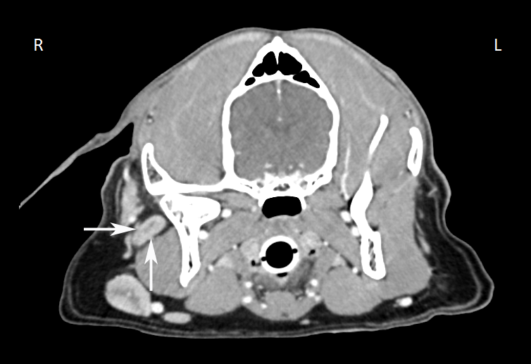
The parotid lymph node (Ln. parotidea) (Figure 13: 1) is found as a large lymph node on each side of the head. It is observed to be between 1.0 to 2.5 cm long, 0.5 to 1.5 cm wide, and 0.4 to 1.0 cm thick. It is located just caudal to the temporomandibular joint and is partially covered by the parotid gland, with a portion protruding rostrally from the parotid gland. This latter portion of the lymph node lies on the caudal margin of the mandible and the M. masseter, where it is covered only by a thin layer of fatty connective tissue, skin muscles, and skin. The lymph node can also be located in the angle formed by the dorsal buccal nerve and the zygomatic branch of the facial nerve. The dorsal buccal nerve is found along the ventral border of the lymph node, while the zygomatic branch of the facial nerve lies adjacent to its caudal border. The superficial temporal nerve emerges at the medial border of the lymph node with branches of the corresponding artery and vein, which are initially medial to the lymph node.
Occasionally, a second small parotid lymph node can be found and may be located dorsally or ventrally from (Figure 14: s’), or on the caudal border of, the main parotid lymph node. This second parotid lymph node was observed 4 times out of 36 cases; once on both the right and left sides, and 3 times on only one side.
A small, sometimes double, lymph node (Figure 14: s”) may be located further caudal to the parotid lymph node and under the parotid gland – it is not a parotid lymph node as reported by Merzdorf [24], but a lateral retropharyngeal lymph node.
The absolute weight of the parotid lymph node on both sides varies between 0.02 and 3.00 g, and the relative weight (relative to the weight of the dog’s body) from 0.0002 to 0.0098%.
Afferent drainage
The parotid lymph node receives afferent lymph vessels from the skin of the caudal half of the bridge of the nose, the forehead, the eyelids, the rostral half of the dorsal aspect of the head, the area of the zygomatic arch and the M. masseter, and the skin of the ear. The parotid lymph node also drains the temporomandibular joint, various head bones (nasal, frontal, parietal, zygomatic, temporal, and mandible) and various muscles of the head (M. zygomaticus, M. temporalis, and M. masseter, facial skin muscles), the outer nose, outer ear (muscles and concha), parotid gland, and the eyelids, lacrimal caruncle, and lacrimal gland.
Efferent drainage
Two to 3 efferent vessels leave the parotid lymph node and either pass between the parotid lymph node and the M. digastricus, or under the M. digastricus, and over both surfaces of the great hyoid branch towards the medial retropharyngeal lymph node. If the lateral retropharyngeal lymph node is present, the efferent vessels of the parotid lymph node will also drain to this lymph node.
Clinical Notes


