Axillary Lymph Nodes
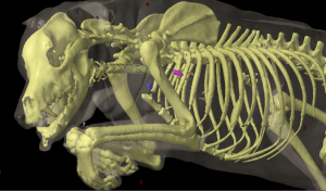
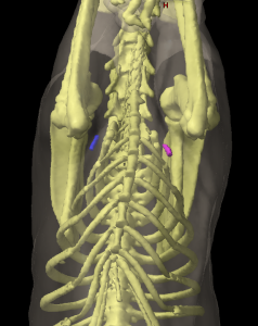
The axillary lymph node (Figures 16: e; 36: d) is almost always a single lymph node; in 29 out of 33 cases I found only 1 lymph node, and in 4 cases there were 2 lymph nodes: 1 lymph node on each side (1 case), 2 on the left side (1 case), and 2 on the right side (2 cases). The length of the main lymph node varied between 0.3 and 5 cm, the width between 0.2 and 5 cm, and the thickness between 0.2 and 1.7 cm. The lymph node usually has an almost round shape and is relatively flattened.
The axillary lymph node lies in abundant fatty tissue on the medial side of the shoulder at the level of the shoulder joint, however, depending on the size of the dog, it may be found 2 to 5 cm caudal from the shoulder joint in the triangle formed by the subscapular and brachial arteries and veins. The lateral surface of the lymph node abuts the M. teres major (Figure 36: 2), and the medial surface of the lymph node abuts the M. pectoralis profundus, but most commonly the M. transversus costarum near the M. scalenus supracostalis at the level of the 1st intercostal space or the 2nd rib (Figure 16: e). The lymph node is therefore enclosed between the M. teres major on one side and the M. transversus costarum and M. pectoralis profundus on the other side. The caudal pectoral nerve, frequently divided, runs over either the medial surface or ventral border of the lymph node, and the thoracodorsal artery and vein run along the dorsal border of the lymph node.
The absolute weight of the lymph node on both sides fluctuated between 0.09 and 36.82 g, and the relative weight between 0.0012% and 0.0695%.
Afferent drainage
The axillary lymph node(s) drain the lymph vessels from the skin of the region of the dorsal, lateral, and ventral thoracic and abdominal wall located between the shoulder muscles and a transverse plane at the level of the last rib, from the skin on the lateral side of the shoulder, the brachium and the olecranon, lymph vessels from the antebrachial fascia, all muscles of the forelimb and their tendons, as well as the M. trapezius, brachiocephalicus, latissimus dorsi and pectoralis profundus, and the abdominal muscles. The axillary lymph node also drains the lymph vessels from nearly all bones of the forelimb (with the exception of the phalanges, and the shoulder, elbow and carpal joints), the mammary gland, and the efferent vessels of the accessory axillary lymph node.
Efferent drainage
Four to 5 efferent vessels emerge from the lymph node, and almost always quickly merge to form a single larger vessel (Figure 16: e). This larger vessel, often forming large dilations, runs cranioventrally over the M. transversus costarum to the 1st rib, where it crosses the medial side of the axillary vein at its origin, and then the lateral side of the initial part of the common jugular vein, often bifurcating into 2 to 3 terminal branches. On the left side, the vessel – or its terminal branches – drain into the thoracic duct or one of its terminal branches, directly into the external jugular vein, or into the end of the left tracheal duct. On the right side, the vessel – or its terminal branches – drain into the end of the right tracheal duct, the right lymphatic trunk or one of its terminal branches, or directly into the end of the external jugular vein. The terminal drainage is therefore highly variable; some of these variations for the left side are shown in Figures 3, 5, and 16: e, and for the right side in Figures 9 and 10: e. If there are 2 axillary lymph nodes (Figures 7: e, e1; 16: e), some of the 2 to 3 efferent vessels of the caudal lymph node open into the cranial lymph node, and some merge with the efferent vessels of the cranial lymph node.
Clinical Notes


