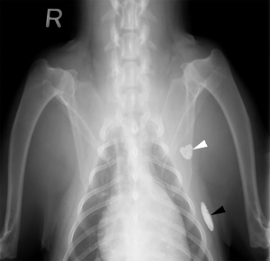
Accessory Axillary Lymph Nodes
The accessory axillary lymph node (Figures 13: 4; 16: e’) is a relatively small, infrequent lymph node. It was found in only 10 of 43 cases: 6 times on both sides, 3 times only on the right, and 1 time only on the left. The lymph node was always found alone. It is located 4 to 8 cm dorsal to the olecranon, just dorsal to the M. pectoralis profundus and just ventral to the M. latissimus dorsi, close to the angle formed between them at the caudal border of the muscles of the shoulder and brachium on the 3rd and 4th ribs, most often in the 3rd intercostal space.
The lymph node is covered only by the skin and the M. cutaneous trunci (Figure 13: m), although in rare cases, it is located on the medial side of the M. latissimus dorsi.
Afferent drainage
The accessory axillary lymph node drains the lymph vessels of the skin on the lateral and ventral thoracic wall and on the caudal aspect of the lateral and medial side of the humerus and olecranon, the lymph vessels from the M. cutaneous trunci, the M. pectoralis profundus, and from the mammary gland. If the accessory axillary lymph node is absent, the afferent vessels from the aforementioned structures drain directly to the axillary lymph node, providing evidence that the accessory axillary lymph node is associated with the axillary lymph node. This is in contrast to Merzdorf [24], who named this lymph node the cubital lymph node. Based on its location, the accessory axillary lymph node in dogs may be compared to the infraspinous lymph node in cattle, although the lymph node in cattle has a completely different drainage region than the lymph node in dogs.
Efferent drainage
The efferent vessels from the accessory axillary lymph node merge to form 2 to 3 vessels (Figure 16: e, e’), which form anastomoses among themselves, and drain into the axillary lymph node.
Clinical Notes


