
Figure 30: Lymph Vessels of the Mammary Gland of the Dog
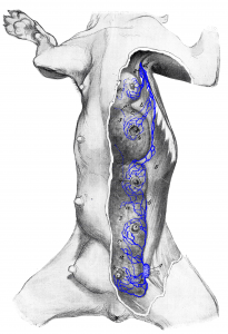
Figure 30: The skin is removed from the left half of the mammary glands. a, a lymph vessels of the parenchyma emerging on the surface; b, b lymph vessels emerging from under the mammary gland; c, c lymph vessels of the skin running deep (the remaining lymph vessels are from the skin and teat); d lymph vessels running to the axillary lymph node; e, e’, e” lymph vessels running deep to the sternal lymph node. 1 superficial inguinal lymph node (slightly pulled out from under the mammary gland); a second superficial inguinal lymph node (1’ ) is covered by the mammary gland; 2 accessory axillary lymph node; 3, 3 mammary gland; 4, 4 teats; 5 M. obliquus abdominis externus. Source: Dr. Hermann Baum (1918). (This work is in the public domain).
