Chapter 13 – Interventional / Vascular (Invasive)
Venous Access
Peripherally Inserted Central Catheter (PICC), Dialysis Catheter, Tunneled Catheter
Case 1
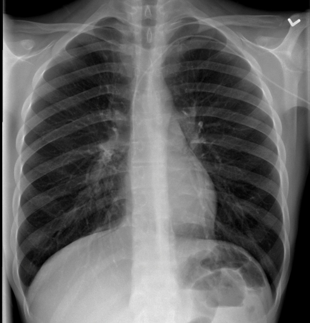
Peripherally Inserted Central Vein Catheter (PICC)
Clinical:
This 55 year old male required intravenous antibiotics for osteomyelitis for 6 weeks. A PICC was inserted. The patient had known COPD.

A small caliber venous catheter was seen entering from the left arm. The tip was at the SVC-Right atrial junction. This was a peripherally inserted central vein catheter (PICC).
Case 2
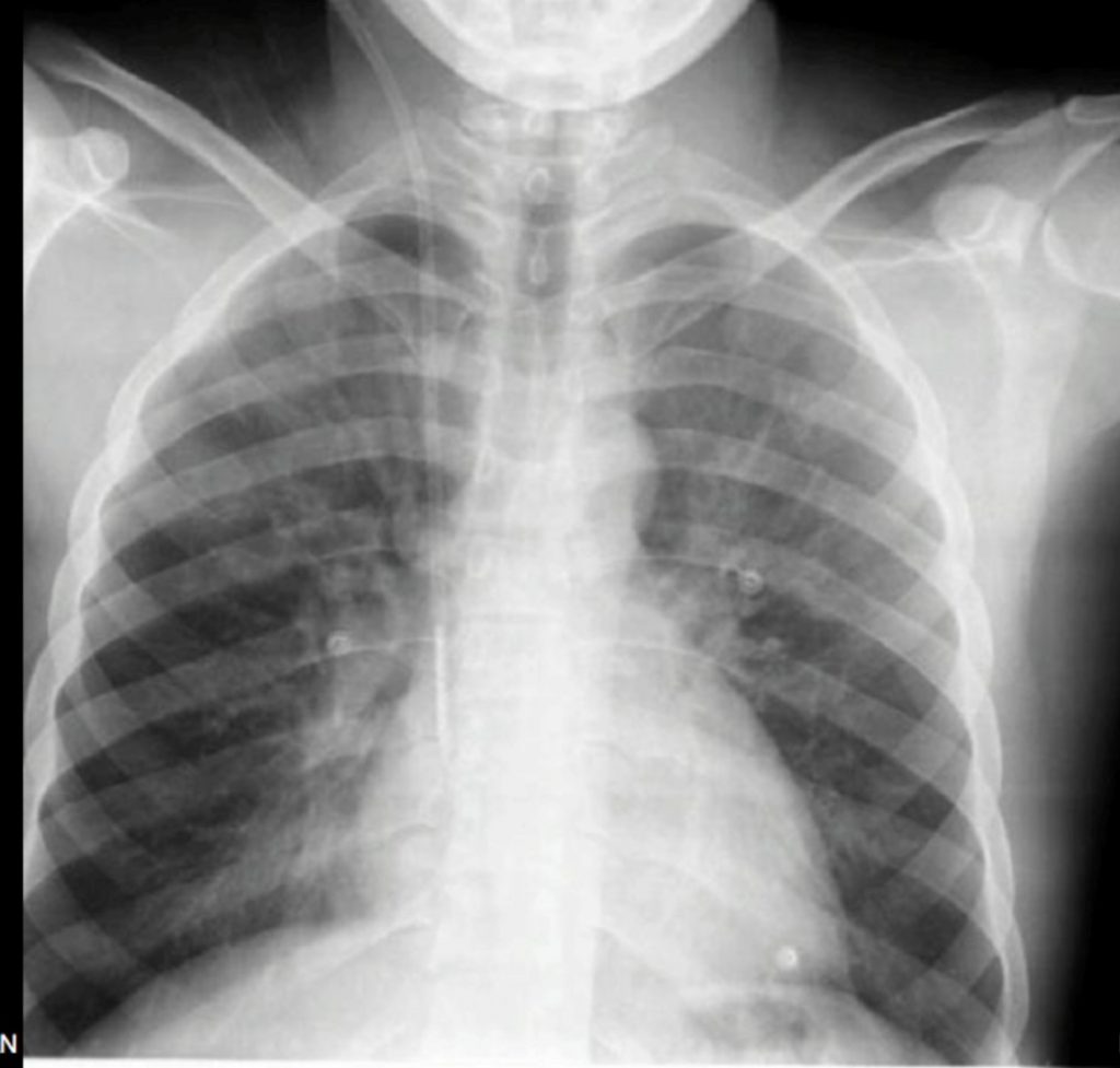
Jugular Catheter
Clinical:
This 45 year old patient required urgent dialysis after developing an immune mediated glomerulonephritis.

A larger caliber catheter was seen. This was a 13F catheter suitable for dialysis. The tip position was satisfactory. This was not a tunneled catheter.
Case 3
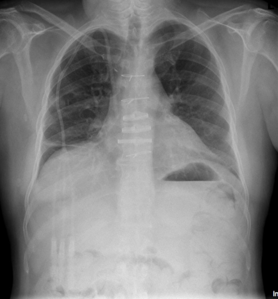
Tunneled Central Line
Clinical:
This 73 year old male required a tunneled line for long-term treatment related to a Stem-Cell Transplant.

A large caliber, triple lumen, catheter was seen superimposed on the upper right chest and neck. The tip of the catheter was at the SVC-Right atrial junction. This was a tunneled catheter implanted for long-term treatment related to a stem cell transplant.
Discussion:
- Peripherally inserted central catheters (PICC),
- Temporary (non-tunneled) central venous catheters,
- Long-term (tunneled) central venous catheters,and
- Implantable ports.
- Peripherally inserted central catheters are essentially long IVs (Case 1). These catheters typically range from 3 to 7French and are inserted in a forearm or upper arm vein. The catheter may have one or two lumens and extends from the puncture site to the superior vena cava. This type of catheter is ideal for administration of intermediate-term medications such as antibiotics or chemotherapy.
- Temporary (non-tunneled) subclavian, femoral, and internal jugular vein catheters are commonly used for medication delivery, central venous pressure monitoring, and short-term hemodialysis (Case 2). Many temporary catheters are constructed of polyurethane. This material is relatively rigid at room temperature but softens when placed in the body. Temporary catheters typically range from 6 to 13French and are most often used for several days to several weeks.
- Long-term (tunneled) catheters are composed of Silastic (silicone elastomer) or polyurethane (Case 3). Silastic is compliant and easily passes through tortuous vessels. Tunneled catheters travel through a short (8 to 15 cm) subcutaneous tunnel before entry into an accessed vein. A polyester cuff on the catheter becomes incorporated into the subcutaneous tissues and helps secure the catheter in place. Although somewhat controversial, the theoretical benefits of the tunneled catheter include decreased risk of infection compared with non-tunneled catheters and decreased risk of inadvertent catheter removal. Unequivocal benefits of tunneled internal jugular vein catheters include improved cosmetic appearance and patient comfort. In general, tunneled catheters are preferred to non-tunneled catheters in patients who require central venous access for longer than 2 weeks.
- Implantable ports consist of a single or dual-lumen reservoir attached to a catheter. The port reservoir is implanted in the arm or chest, and the catheter is tunneled to the accessed vein. These devices are typically used for long-term, intermittent, venous access such as that required for chemotherapy. The port is accessed using a Huber (non-coring) needle. Some radiologists prefer chest implantation of these devices while other radiologists prefer arm ports, particularly in young women because of better cosmetic appearance. Among central venous catheters, ports have the lowest incidence of infection because they are implanted completely beneath the skin.
Attributions
Figure 13.5 Chest X-ray of a PICC line by Dr. Brent Burbridge MD, FRCPC, University Medical Imaging Consultants, College of Medicine, University of Saskatchewan is used under a CC-BY-NC-SA 4.0 license.
Figure 13.6 Chest X-ray of a Jugular Catheter by Dr. Brent Burbridge MD, FRCPC, University Medical Imaging Consultants, College of Medicine, University of Saskatchewan is used under a CC-BY-NC-SA 4.0 license.
Figure 13.7 Chest X-ray of a Tunneled Catheter by Dr. Brent Burbridge MD, FRCPC, University Medical Imaging Consultants, College of Medicine, University of Saskatchewan is used under a CC-BY-NC-SA 4.0 license.

