
Chapter 7 – Breast
Introduction to Breast Imaging
Communicate the basic technical aspects of mammography, and breast ultrasound
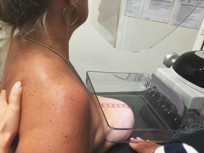
Mammograms are x-ray images of the breast tissue obtained in a standardized manner following a routine, reproducible, breast positioning strategy and a stringent quality assurance program. The mammography machine acquires images while the breast tissue is being firmly compressed by plastic paddles. The compression spreads the breast tissue evenly and minimizes the thickness of the tissue to be imaged. Spreading and thinning the breast tissue results in the acquisition of images using less radiation as there is less tissue thickness for the x-rays to pass through. The x-rays generated by the machine are based upon a molybdenum, rather than a tungsten, x-ray tube anode which optimizes the x-ray beam for imaging soft tissues (fat, connective and mammary tissue).
Ultrasound uses traditional probes with higher frequency to better image the breast tissue that is in close proximity to the overlying skin.
Mammography/Breast Imaging Centres in Canada are accredited and audited for quality by the Canada Association of Radiologists.
Mammography Image Acquisition
Canadian Association of Radiologists – Mammography Accreditation – http://www.car.ca/en/accreditation/map.aspx
Breast Cancer Screening
Screening imaging for the detection of breast cancer was established in an attempt to image tumours prior to their development into clinically detectable abnormalities.
Imaging:
There are a variety of available guidelines that facilitate the development of a physician-patient strategy for screening for breast malignancy. These strategies may include clinical, laboratory, and imaging parameters. These guidelines have been refined to address unique patient sub-groups i.e. low risk, average risk, intermediate risk, and high risk of developing breast cancer, There are a variety of Guidelines for Breast Screening.
Canadian Association of Radiology (CAR)
Practice Guidelines
- Asymptomatic women 40 – 49 years should undergo screening mammography every year
- Asymptomatic women aged 50 – 75 should undergo screening mammography every one to two years
- Women over the age of 74 should have screening mammography at one to two-year intervals if they are in good general health
Normal Mammogram Cranial-Caudal (CC) Projection

Normal Mammogram Medio-Lateral Oblique (MLO) Projection


Note how the above figures (Figure 7.2 and Figure 7.3) contrast from the following figure (Figure 7.4) which shows a breast mass that can be visualized both on mammography and ultrasound.
Breast Mass Mammography

Breast Mass Ultrasound

For More Information:
CAR: Practice Guidelines and Technical Standards for Breast Imaging and Intervention
http://www.car.ca/uploads/standards%20guidelines/car_breastimagingguidelines_2016_en.pdf
ACR: Appropriateness Criteria – Breast Imaging – https://acsearch.acr.org/docs/70910/Narrative/
Attributions
Figure 7.1 Acquisition of a mammogram in the cranial-caudal projection. Image courtesy of, http://lifeloveandhiccups.blogspot.ca/search?q=mammogram . Used with permission.
Figure 7.2A Normal Mammography Image, CC Projection by Dr. Brent Burbridge MD, FRCPC, University Medical Imaging Consultants, College of Medicine, University of Saskatchewan is used under a CC-BY-NC-SA 4.0 license.
Figure 7.2B Normal Mammography Image, MLO projection by Dr. Brent Burbridge MD, FRCPC, University Medical Imaging Consultants, College of Medicine, University of Saskatchewan is used under a CC-BY-NC-SA 4.0 license.
Figure 7.3 Normal Breast Ultrasound by Dr. Brent Burbridge MD, FRCPC, University Medical Imaging Consultants, College of Medicine, University of Saskatchewan is used under a CC-BY-NC-SA 4.0 license.
Figure 7.4A Mammography of a breast mass by Dr. Brent Burbridge MD, FRCPC, University Medical Imaging Consultants, College of Medicine, University of Saskatchewan is used under a CC-BY-NC-SA 4.0 license.
Figure 7.4B Ultrasound of a breast mass by Dr. Brent Burbridge MD, FRCPC, University Medical Imaging Consultants, College of Medicine, University of Saskatchewan is used under a CC-BY-NC-SA 4.0 license.
