Chapter 15 – Pediatric
Tumors Unique to Children – Wilms Tumour
Case
Tumors Unique to Children, Wilms Tumor
Clinical:
History – Failure to thrive. Enlarging abdominal girth in a 3 year old male.
Symptoms – Lethargic. Enlarging abdomen.
Physical – The abdomen was distended and firm. Palpation of the abdomen was difficult due to the severe distention but there was suspicion for an extremely large mass in the left upper quadrant/left mid abdomen.
DDx:
Adrenal mass
Renal mass
Mesenteric mass
Pancreatic mass
Imaging Recommendation
X-rays, chest and abdomen
Ultrasound.
CT and/or MR for further characterization and staging.


Imaging Assessment
Findings:
Chest and abdomen x-rays
The chest x-ray was normal. There was a large mass in the left upper abdomen that obscured the fat plane between the kidney and the spleen. The mass pushed the stomach and the colon to the right. The abdomen was distended.
Ultrasound
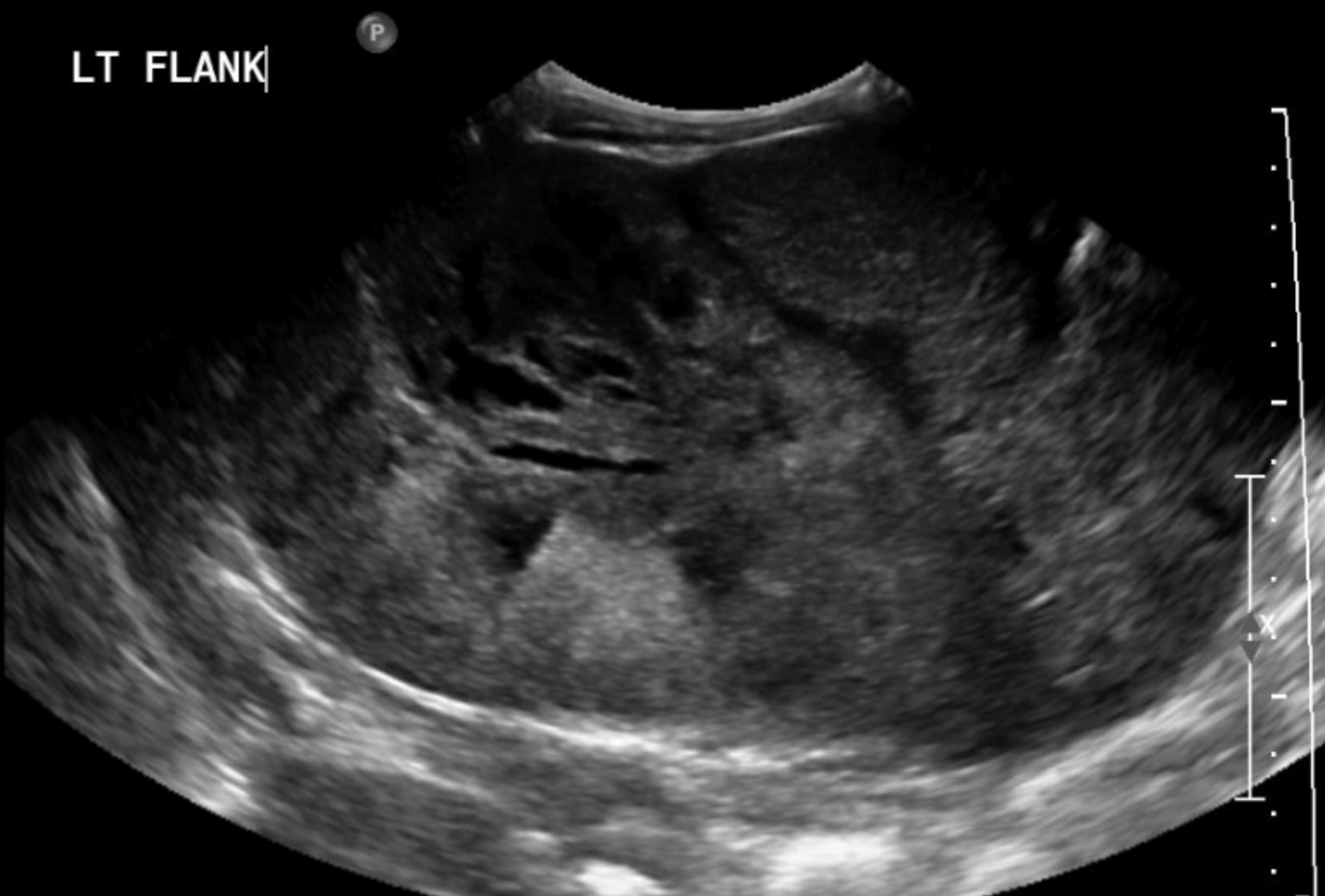
There was a reniform shaped mass in the left upper abdomen with areas of low attenuation throughout the mass.
Computed Tomography
Multiple lung nodules were seen. There was a mixed density mass in the left upper abdomen that was pushing a small amount of normal renal parenchyma posteriorly. The renal vein could not be visualized. No obvious adenopathy.
Interpretation:
Large mass arising from the left kidney. Stage 4, metastatic lung nodules.
Diagnosis:
Wilms tumor of the left kidney – Pathology proven
Discussion:
Wilms tumor (WT), also known as nephroblastoma, is an embryonal renal neoplasm consisting of metanephric blastema; it accounts for 85% of cases. It represents 5.9% of all pediatric malignant tumors and has an annual incidence of 7.6 cases/million children younger than 15 years. The peak incidence occurs at 2 to 3 years. Of all patients, 13% can present with bilateral tumors.
Clinical Presentation
WT is typically discovered incidentally during a physical examination or because parents palpate an abdominal mass. Other presenting symptoms include abdominal pain and hematuria, which may signify tumor invasion into the collecting system or ureter. Another 25% develop hypertension, which is thought to occur secondary to disturbances in the renin-angiotensin feedback loop. Less than 10% of patients have atypical presentations.
Imaging findings may include:
- X-rays of the abdomen may reveal a large mass. This mass may displace the stomach or colon. The fat margin between the spleen and the liver may be obscured by the mass.
- Ultrasonography is initially performed to determine whether the tumor is of renal origin, is cystic or solid, or extends into the renal vein or IVC.
- A CT scan is useful to delineate Wilms tumor from neuroblastoma and also to evaluate for regional adenopathy, contralateral kidney involvement, and metastatic disease.
- MRI is a useful adjunct for evaluating intravascular invasion; however, an ultrasound study may be preferred.
- Lung metastases, which are present in 8% at the time of diagnosis, can be identified on an initial chest radiograph, but CT scan is obtained routinely and will be more sensitive.
Pathology
- The histology of Wilms tumor is categorized as favorable or unfavorable. Favorable histology is more common, characterized by the presence of three elements—blastemal, stromal, and epithelial cells.
- Wilms tumor with predominantly epithelial differentiation behaves less aggressively and tends to be stage I when it is diagnosed early.
- Blastemal-predominant tumors tend to be clinically aggressive and are associated with advanced disease.
- Outcomes are correlated with histopathologic features and tumor stage. Unfavorable histology is defined by the presence of anaplasia, clear cell sarcoma, or rhabdoid tumor.
Staging
The International Society of Pediatric Oncology (SIOP) staging system is based on pre-operative chemotherapy but is applied after resection. The presence of metastases is evaluated at presentation, relying on imaging studies, and chemotherapy is instituted before operative intervention. The National Wilms Tumor Study Group (NWTSG) has also developed a staging system that incorporates the clinical, surgical, and pathologic information that was obtained at the time of resection but stratifies patients before the initiation of chemotherapy. The advantage of this system is that it favors stage-based therapy, thereby avoiding unnecessary chemotherapy in patients who might not otherwise benefit from it.
Attributions
Figure 15.9A Abdominal ultrasound of the left flank/renal region demonstrating a mass by Dr. Brent Burbridge MD, FRCPC, University Medical Imaging Consultants, College of Medicine, University of Saskatchewan is used under a CC-BY-NC-SA 4.0 license.
Figure 15.9B Axial CT of the abdomen revealing a very large mass by Dr. Brent Burbridge MD, FRCPC, University Medical Imaging Consultants, College of Medicine, University of Saskatchewan is used under a CC-BY-NC-SA 4.0 license.

