Chapter 16 – Urogenital
Testicular Torsion
ACR – Urologic – Acute Scrotal Pain, Without Trauma
Case
Testicular Torsion
Clinical:
History – This 15 year old male presented with severe right scrotal pain.
Symptoms – Severe right scrotal pain.
Physical – The right testicle was mildly enlarged and very tender.
DDx:
Epididymitis
Orchitis
Testicular torsion
Imaging Recommendation
ACR – Urologic – Acute Scrotal Pain, Without Trauma, Variant 1
Doppler Ultrasound of the Scrotum


Imaging Assessment
Findings:
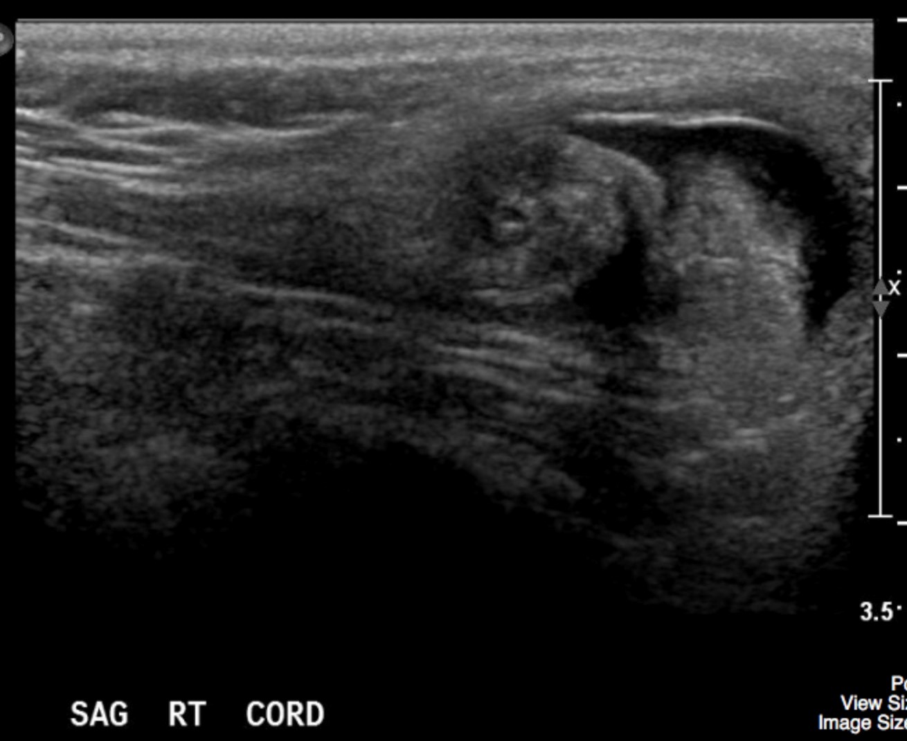
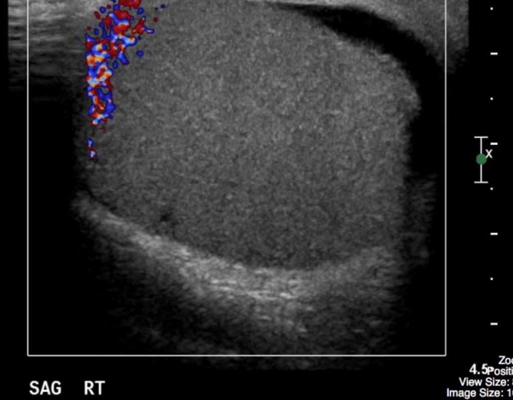
The right testicle was uniform in appearance. There was no Doppler flow to the right testicle. A ‘whirlpool’ appearance to the spermatic cord was visualized.
Interpretation:
Right testicular torsion
Diagnosis:
Testicular torsion
Discussion:
The ability to confidently establish a surgical versus a nonsurgical diagnosis for acute scrotal pain is important. The benefits of early surgery for testicular salvage in ischemic disease, primarily torsion of the spermatic cord, are well known, but must be balanced against the costs of operating unnecessarily on a large number of patients with nonsurgical disease, primarily acute epididymitis.
The most common differential diagnoses of the acute scrotum include 1) torsion of the spermatic cord, 2) torsion of the testicular appendages, and 3) acute epididymitis or epididymoorchitis. Less common diagnoses include strangulated hernia, segmental testicular infarction, trauma, testicular tumor, and idiopathic scrotal edema.
Acute epididymitis is commonly the cause of acute scrotal pain in adults and should be differentiated from testicular torsion. Testicular torsion is rare in patients older than age 35. Acute epididymitis is commonly the cause of acute scrotal pain in patients younger than age 18, very common in patients age 19 to 25, and overwhelmingly the etiology in patients older than age 25.
Acute scrotal pain in prepubertal boys occurs most commonly from torsion of the testicular appendages, a process that may clinically mimic testicular torsion or epididymo-orchitis.
Patients with testicular torsion typically present with abrupt scrotal pain, whereas those with epididymitis have a more gradual onset of pain. Patients with torsion will have a normal urinalysis, whereas those adults (but not children) with epididymitis will have an abnormal urinalysis. There is, however, overlap in the clinical presentation of the different causes of acute scrotal pain.
Imaging in clinically equivocal cases may lead to an early diagnosis of testicular torsion and thus decrease the number of unnecessary surgeries.
US findings may include:
- Grayscale US is most sensitive to the earliest changes resulting from decreased or absent testicular perfusion.
- In patients with torsion, however, a normal homogenous echo pattern is likely to indicate a viable testis, whereas a hypoechoic or inhomogeneous testis is likely to be nonviable.
- The finding of a twisted cord has been referred to as the “whirlpool sign” and can be found at the external inguinal ring, above the testis, and posterior to the testis.
- Color Doppler US (CDU) is a valuable examination for evaluating testicular perfusion.
- Color duplex Doppler involves the simultaneous acquisition and display of color Doppler and spectral Doppler waveforms in conjunction with grayscale sonographic imaging. Settings optimized to detect slow flow include use of a small color-sampling box, lowest pulse repetition frequency, and lowest possible threshold.
- CDU is readily available and can be done quickly without any specific preparation.
Attributions
Figure 16.5A Sagittal ultrasound of right spermatic cord demonstrating the “whirlpool sign”, by Dr. Brent Burbridge MD, FRCPC, University Medical Imaging Consultants, College of Medicine, University of Saskatchewan is used under a CC-BY-NC-SA 4.0 license.
Figure 16.5B Sagittal doppler ultrasound of right testicle revealed no detectable blood flow in the testicle by Dr. Brent Burbridge MD, FRCPC, University Medical Imaging Consultants, College of Medicine, University of Saskatchewan is used under a CC-BY-NC-SA 4.0 license.

