Chapter 12 – Head and Neck
Retropharyngeal Abscess – Child
Case
Retropharyngeal Abscess
Clinical:
History – Acute malaise, fever, and lethargy.
Symptoms – Trouble swallowing.
Physical – Fever. The neck was tender to palpation. No masses palpable. The tonsillar fossae were reddened with no purulent exudate present. The neck was stiff and neck range of motion was painful.
Laboratory – WBC is elevated.
DDx:
Tonsillitis
Pharyngitis
Epiglottitis
Foreign Body Ingestion
Retropharyngeal abscess
Imaging Recommendation
Soft tissue x-rays of the neck

Imaging Assessment
X-Rays:
Findings:
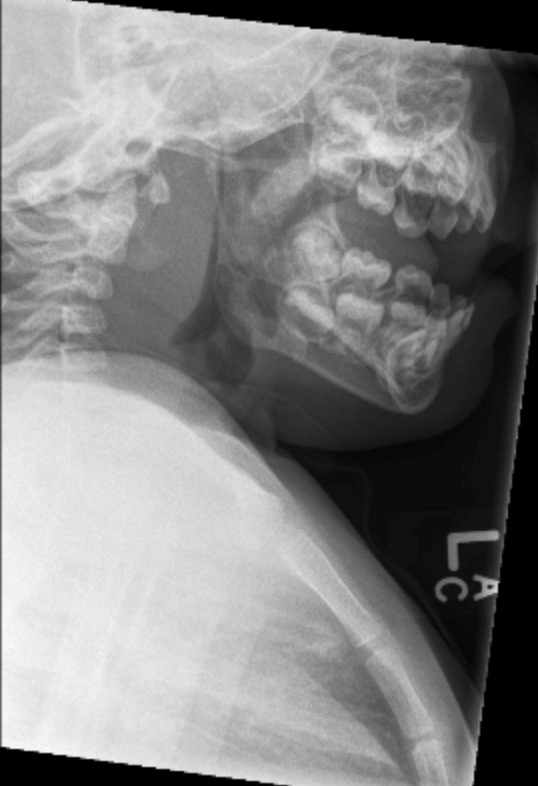
The pre-vertebral soft tissues were massively, uniformly swollen.
Interpretation:
Probable pre-vertebral/retropharyngeal abscess. Emergent CT was recommended.
Computed Tomography:
Findings:
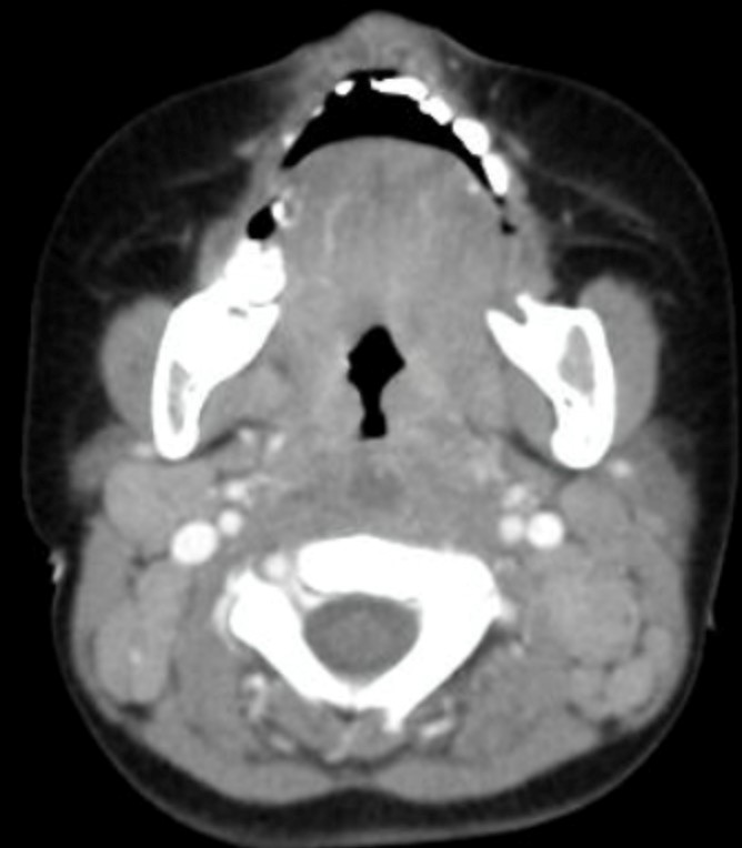
Images were acquired with intravenous contrast. The pre-vertebral soft tissues were thickened. There was a central low density area in the thickened tissue. This low density area demonstrated rim enhancement with intravenous contrast. There was scalloping of the wall of the low density tissue.
Interpretation:
Retropharyngeal abscess.
Diagnosis:
Retropharyngeal abscess.
Discussion:
Retropharyngeal and lateral pharyngeal infections are most often polymicrobial; the usual pathogens include group A streptococcus, oropharyngeal anaerobic bacteria, and Staphylococcus aureus. In children younger than age 2 yrs., there has been an increase in the incidence of retropharyngeal abscess, particularly with S. aureus, including methicillin-resistant strains. Mediastinitis may be identified on CT in some of these patients. Other pathogens can include Haemophilus influenzae, Klebsiella, and Mycobacterium avium-intracellulare.
Imaging findings include:
- Soft-tissue neck films taken during inspiration with the neck extended might show increased width of the pre-vertebral soft tissue and/or an air–fluid level in the retropharyngeal space.
- CT with contrast medium enhancement can reveal central lucency, ring enhancement, or scalloping of the walls of a lymph node.
- Scalloping of the wall of a lymph node or of the fluid containing space, is thought to be a late finding and predicts abscess formation.
Attributions
Figure 12.6A Lateral x-ray of the neck demonstrating pre-vertebral soft tissue swelling by Dr. Brent Burbridge MD, FRCPC, University Medical Imaging Consultants, College of Medicine, University of Saskatchewan is used under a CC-BY-NC-SA 4.0 license.
Figure 12.6B Axial CT Scan of the Head and Neck revealing a retropharyngeal abscess by Dr. Brent Burbridge MD, FRCPC, University Medical Imaging Consultants, College of Medicine, University of Saskatchewan is used under a CC-BY-NC-SA 4.0 license.


