Chapter 17 – Normal, Reference Images, Unlabelled and Labelled

Pelvis

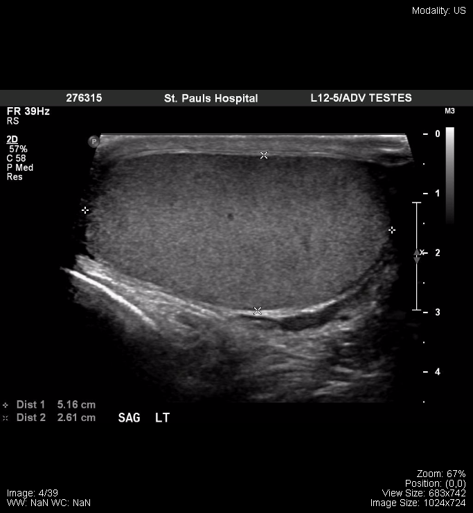
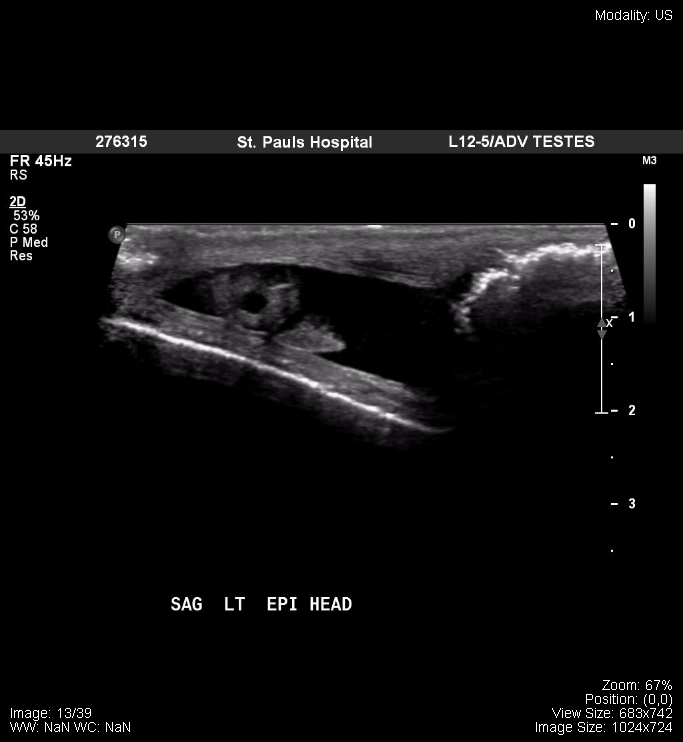
The following is a normal ultrasound of the testicles:
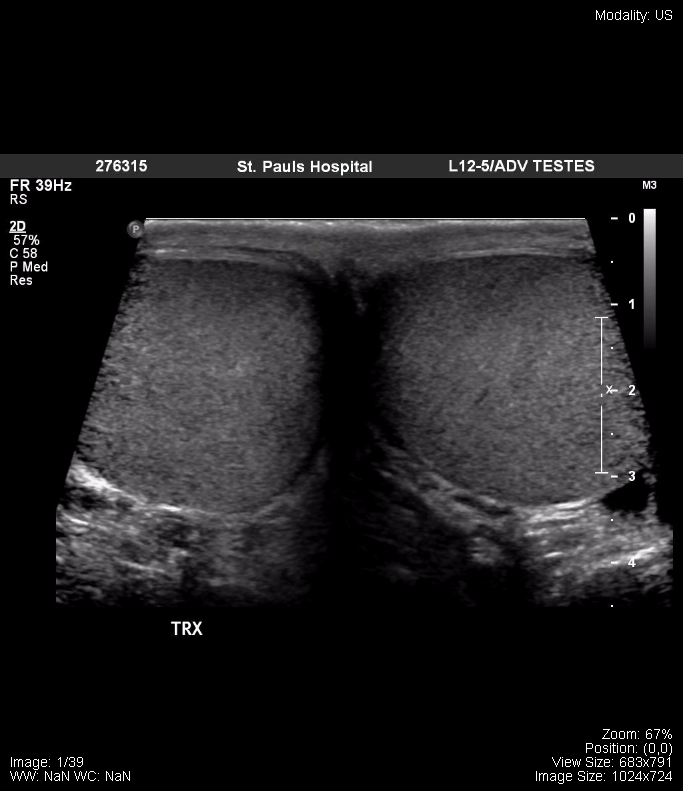
Transverse – both testicles


Sagittal view, left testicle, with measurements


Sagittal view, left epididymis, head


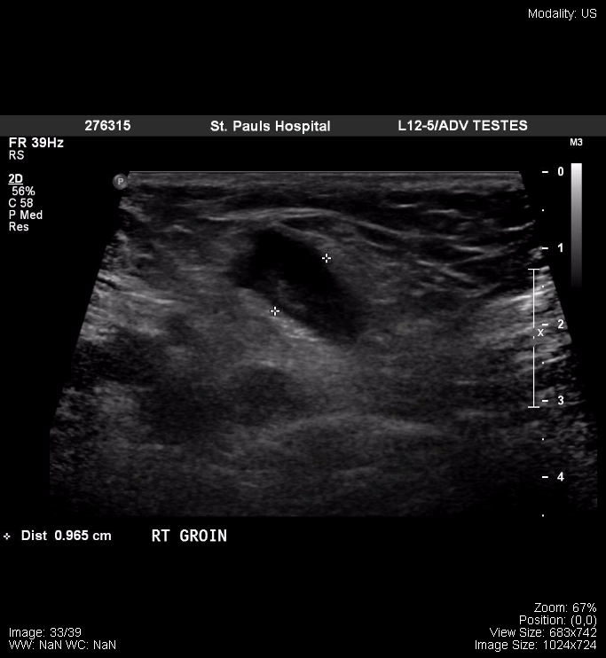
Right Groin


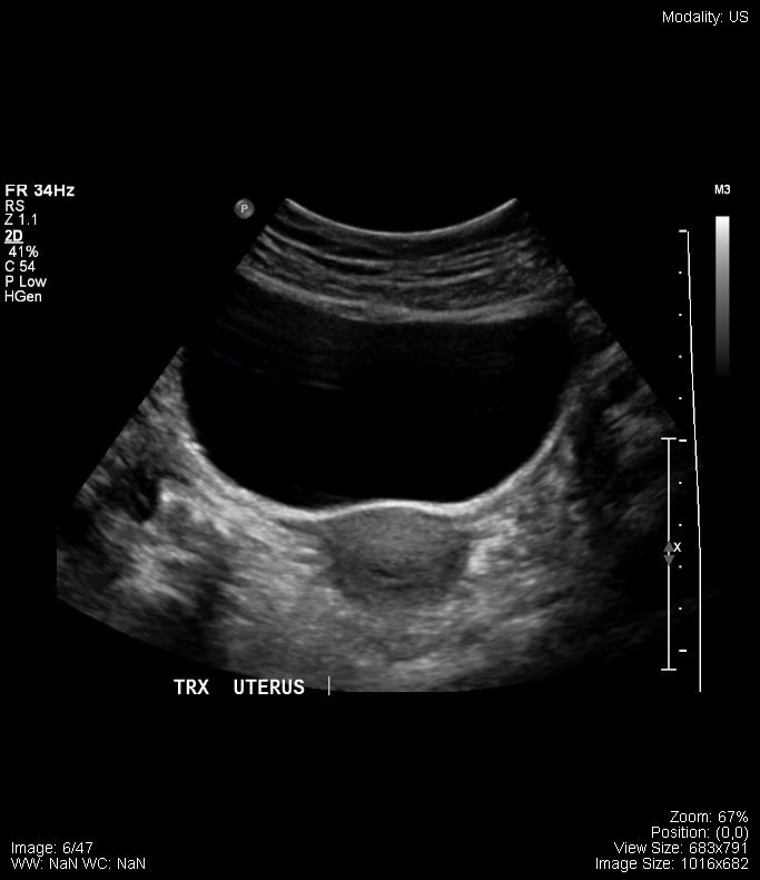
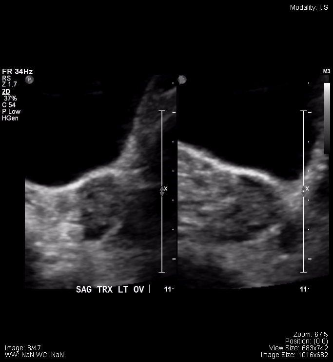
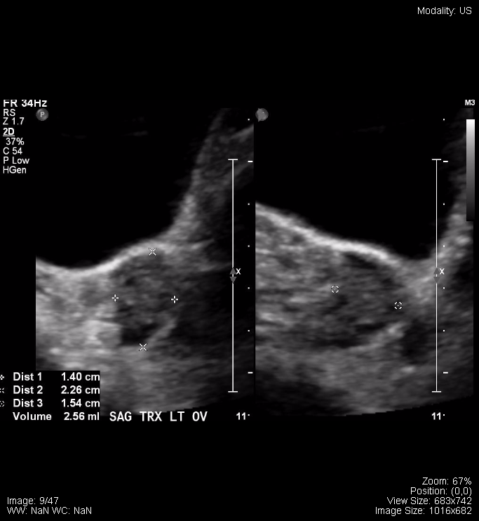
The following are from a normal female pelvic ultrasound:
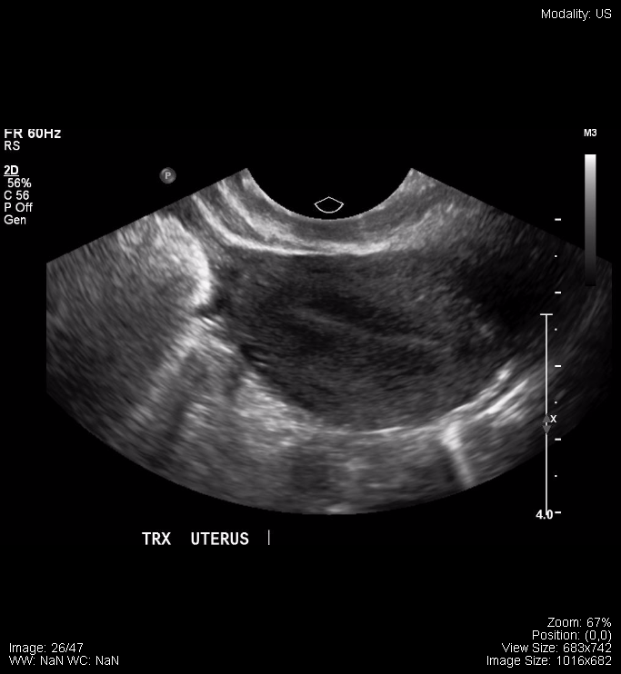
Transverse Uterus

Transverse Uterus 2

Sagittal Ovary

Sagittal Ovary, with measurements

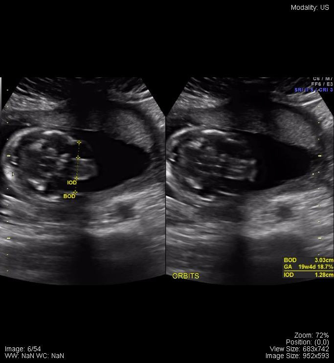
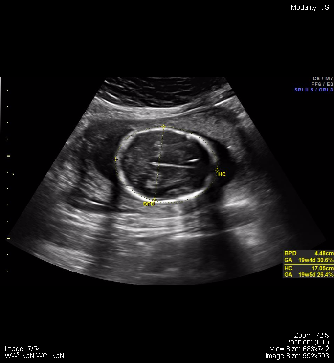
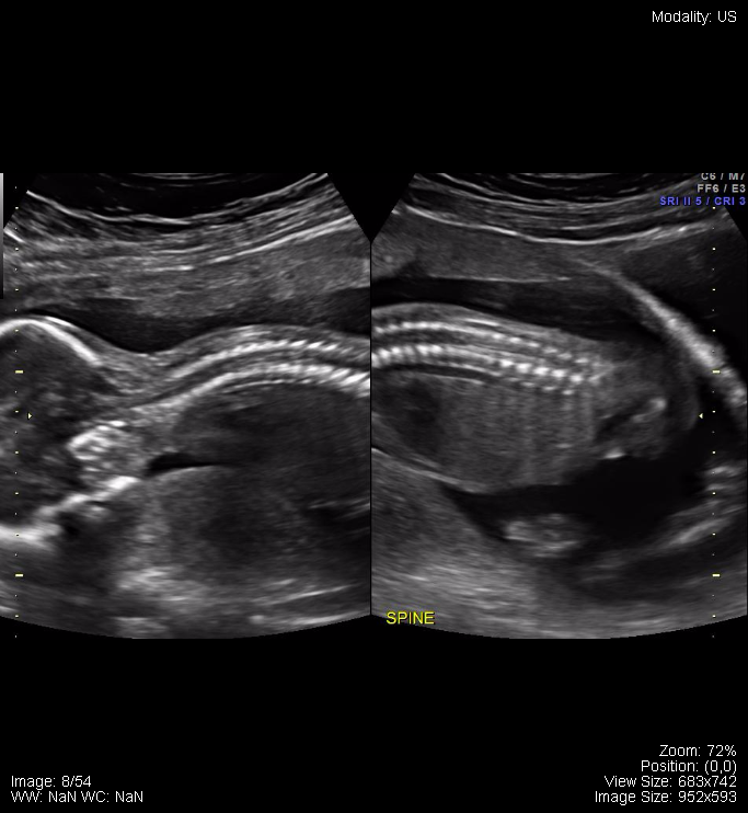
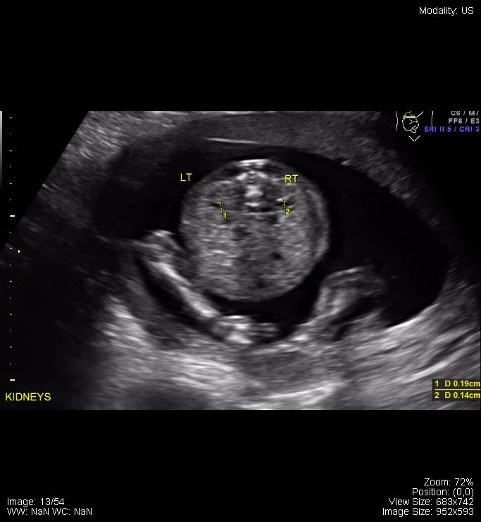
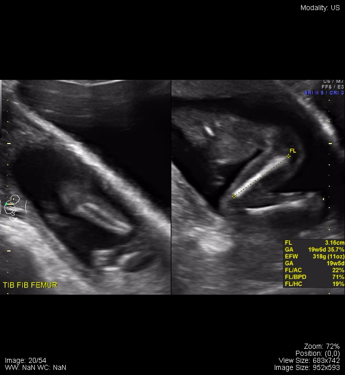
The following are samples from a normal pregnancy ultrasound:
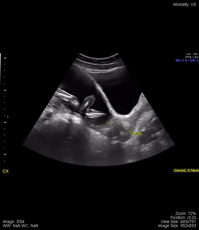
Pregnancy Ultrasound


Fetal Orbits with measurements


Fetal Head with measurements


Fetal spine with measurements


Fetal Kidneys with measurements


Fetal Legs with femur measurements


Attributions

All figures in “Chapter 17: Pelvis” by Dr. Brent Burbridge MD, FRCPC, University Medical Imaging Consultants, College of Medicine, University of Saskatchewan is used under a CC-BY-NC-SA 4.0 license.

License

Icon for the Creative Commons Attribution-NonCommercial-ShareAlike 4.0 International License

Undergraduate Diagnostic Imaging Fundamentals Copyright © 2017 by Brent Burbridge is licensed under a Creative Commons Attribution-NonCommercial-ShareAlike 4.0 International License, except where otherwise noted.