Chapter 3 – Principles of Imaging Techniques
Nuclear Medicine
The patient becomes the source of the radiation required to produce nuclear medicine images after being injected with a radioactive pharmaceutical agent. The pharmacologic agent, bound to the radioactive substance, will concentrate in different organs and tissues based upon the physiology and pharmaco-distribution of the compound administered. The radiation dose administered to the patient is adjusted for the size and weight of the patient. The pharmaceutical agents used to bind and deliver the radioactive isotope must be sterile and quality control must ensure the molecular structure of the drug is stable and effective.
For example, the radioactive substance for a bone scan, Technetium 99m (99mTc), is chemically bound to a pharmacologic agent, diphosphonate , and this is injected intravenously. Technetium is a Gamma-emitter and this radioactivity is emitted as the radionuclide decays. The detector system registers the emitted photons and transforms the energy emitted into pixels on an image display. Therefore, a nuclear medicine bone scan depicts radioactivity arising from bone as the the pharmacologic agent, diphosphonate, physiologically interacts with living bone. Similarly, a renal Nuclear Medicine scan depicts the kidneys, ureters and bladder as the pharmacologic agent is preferentially metabolized and concentrated in these tissues.
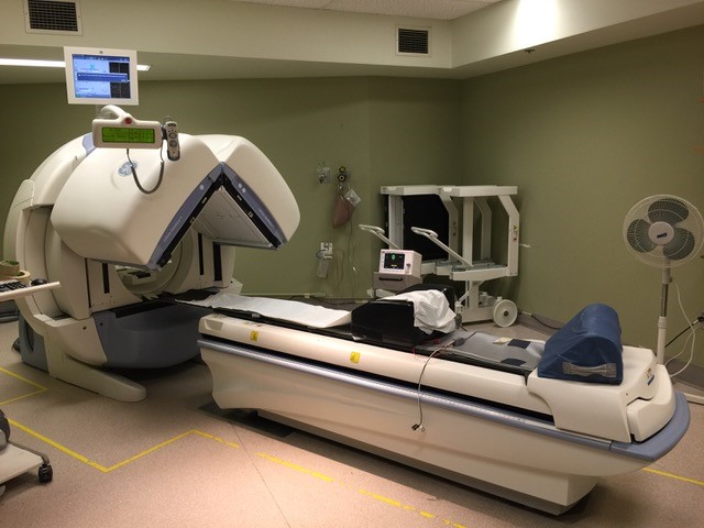
Figure 3.43 depicts a Nuclear Medicine machine. After the patient is injected with the radioactive pharmaceutical they lie on the table in a supine position while the machine detects the emitted radiation.
An example of a normal bone scan for a pediatric patient is provided in Figure 3.44A, while the normal bone scan for an adult is in Figure 3.44B.
Pediatric Bone Scan
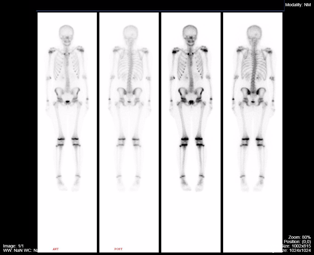
Normal Adult Bone Scan
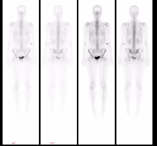
The images above depict a normal pediatric bone scan. Areas of active bone growth i.e. epiphyseal growth plates, demonstrate more radioactivity as the pharmacologic substrate bound to the radioactive Technetium concentrates in areas of heightened bone metabolic activity. This is in contrast to Figure 3.44B, where the adult does not have the same areas of uptake of the Technetium-diphosphonate.
Due to the physics of the detection of emitted photons, the images acquired are low in anatomic detail (spatial resolution), but provide information about the physiology of the tissue being imaged related to the quantitative uptake of the radioactivity into the target tissues. Therefore, regions of the patient’s anatomy that harbor a larger concentration of the injected radioactive substance emit the largest amount of detectable radiation. The areas emitting radiation are displayed in black on static, standard, nuclear medicine images. More detected radiation results in a blacker region of the resulting image. For the pediatric bone scan displayed previously one can appreciate that the growth centres of the child’s bones are more metabolically active and are blacker on the bone scan.
Nuclear Medicine:
- Uses an internal source of radiation (e.g. Technetium 99m)
- Low spatial resolution
- Provides information about the physiology of the tissue being assessed
PET/CT
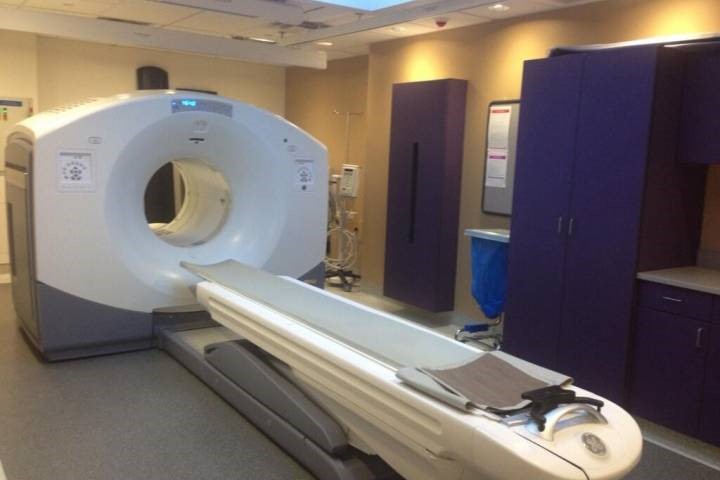
Positron Emission Tomography – Computed Tomography (PET-CT) is a fused imaging technology that detects positron emission from the radioactive source (Fluorine) and creates images for display. There is a CT scanner built into the same apparatus that houses the positron detector and the two examinations can be acquired with the patient remaining in the same anatomic position. The patient lies supine on the table while the table-top moves into the scanner housing. Since the CT and the PET scan are obtained simultaneously it is subsequently possible to overlay (fuse) one image set on the other to correlate the activity level of positron emission with the anatomy seen on the CT. Figure 3.45 illustrates the appearance of a PET/CT scanner.
PET-CT has emerged as a very important imaging tool for the detection of metabolically active tissue. The most common radioactive substance used for PET-CT is fluorine bound to glucose. Elevated tissue metabolic activity for glucose will result in more of the radioactive fluorine concentrating in those tissues. Malignancies and infections metabolize glucose at an accelerated rate and can be imaged with PET even when the CT scan, taken for correlation, is normal.
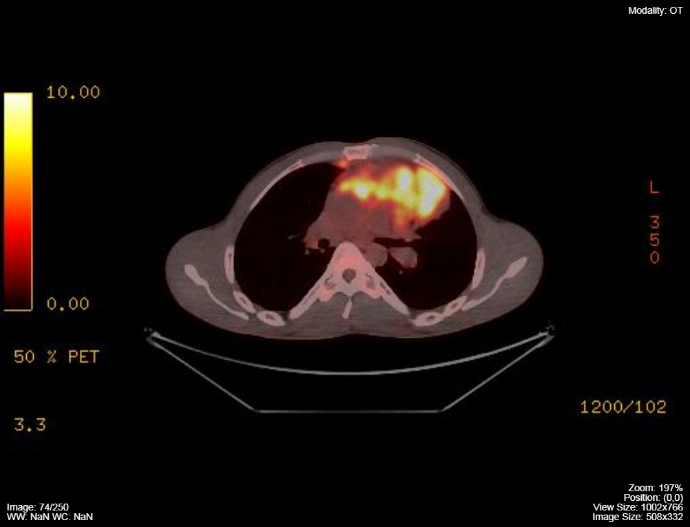
The regions of the fused PET/CT axial scans that have the most metabolic activity are registered as a bright red/orange colour while those areas with little metabolic activity are black. A PET/CT axial scan for a patient with widespread lymphoma and is provided for review in Figure 3.46A.
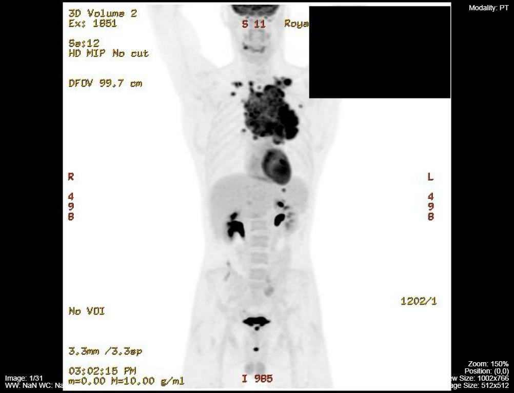
A PET whole body image is displayed in Figure 3.46B. The regions of greater metabolic activity results in a blacker pixels for the PET portion of the scan.
Fused PET/CT
Whole Body PET Image
Attributions
Fig 3.43 Nuclear Medicine Scanner by Dr. Brent Burbridge MD, FRCPC, University Medical Imaging Consultants, College of Medicine, University of Saskatchewan is used under a CC-BY-NC-SA 4.0 license.
Fig 3.44A Normal, Pediatric Nuclear Medicine Bone Scan by Dr. Brent Burbridge MD, FRCPC, University Medical Imaging Consultants, College of Medicine, University of Saskatchewan is used under a CC-BY-NC-SA 4.0 license.
Fig 3.44B Normal, Adult Nuclear Medicine Bone Scan by Dr. Brent Burbridge MD, FRCPC, University Medical Imaging Consultants, College of Medicine, University of Saskatchewan is used under a CC-BY-NC-SA 4.0 license.
Fib 3.45 PET/CT Scanner by Dr. Brent Burbridge MD, FRCPC, University Medical Imaging Consultants, College of Medicine, University of Saskatchewan is used under a CC-BY-NC-SA 4.0 license.
Fig 3.46A PET/CT image of the chest for a patient with lymphoma by Dr. Brent Burbridge MD, FRCPC, University Medical Imaging Consultants, College of Medicine, University of Saskatchewan is used under a CC-BY-NC-SA 4.0 license.
Fig 3.46B PET image of the whole body for a patient with lymphoma by Dr. Brent Burbridge MD, FRCPC, University Medical Imaging Consultants, College of Medicine, University of Saskatchewan is used under a CC-BY-NC-SA 4.0 license.

