Chapter 9 – Chest
Multiple Lung Nodules
Case
Multiple Pulmonary Nodules
Clinical:
History – This 64 year old female had a nephrectomy for renal cell carcinoma 1 year ago. She was lost to follow-up. She was seeing her primary physician for mild chest pain, shortness of breath, and fatigue.
Symptoms – Mild, dull, chest pain. Weight loss (7 kg in 3 months). Fatigue.
Physical – Nephrectomy scar was seen. Nil else.
DDx:
Infection, Pneumonia
Malignancy
Imaging Recommendation
Chest X-ray

Imaging Assessment
Findings:
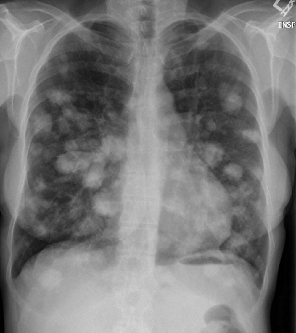
There were multiple nodules in both lungs of varying sizes. They were round and well marginated. No evidence of central necrosis. No evidence of lymphadenopathy. No other findings.
Interpretation:
Embolic infection
Embolic malignancy
Diagnosis:
Metastatic Disease – Renal Cell Carcinoma
Discussion:
Multiple nodules in the lung are most often metastatic lesions that have traveled through the bloodstream from a distant primary (hematogenous spread). Hematogenous spread of infection may also be possible. Multiple metastatic nodules are usually of differing sizes, varying from micronodular to “cannonball” masses, indicating tumour embolization that occurred at different times. They are frequently sharply marginated.
| Possible Tumours of Origin: | |
| Males | Females |
| Colorectal carcinoma | Breast cancer |
| Renal cell carcinoma | Colorectal carcinoma |
| Head and neck tumors | Renal cell carcinoma |
| Testicular and bladder carcinoma | Cervical or endometrial carcinoma |
| Malignant melanoma | Malignant melanoma |
| Sarcomas | Sarcomas |
Possible Primary Tumours that may result in Lung Metastases, by Gender
X-ray findings may include:
- Nodules of similar or varying sizes.
- The patient may be cachectic due to malignancy.
- The nodules may be smooth or lobulated.
- There may be cavitation in some types of nodules or masses.
Attributions
Figure 9.25 Chest x-ray displaying multiple lung nodules by Dr. Brent Burbridge MD, FRCPC, University Medical Imaging Consultants, College of Medicine, University of Saskatchewan is used under a CC-BY-NC-SA 4.0 license.

