Chapter 9 – Chest
Lobar and Lung Collapse – Suspected Lung Malignancy
ACR – Chest – Acute Respiratory Illness in Immunocompetent Patient
Case
Left Upper Lobe Collapse, Central LUL Lung Mass
Clinical:
History – This 58 year old female had a 30 pack/year cigarette smoking history. She had been previously diagnosed with mild emphysema and chronic bronchitis.
Symptoms – She complained of a worsening cough with blood tinged sputum. She also had mild, dull, left chest pain that was persistent. The shortness of breath associated with her emphysema seemed to be slightly worse.
Physical – The trachea was deviated to the left. There were transmitted breath sounds in the left upper chest. There were coarse rales and rhonchi in the left chest. Percussion reveals that the left hemidiaphragm was elevated in comparison to the left. No palpable masses or lymph nodes.
DDx:
Emphysema/Chronic Bronchitis exacerbation
Lobar Collapse due to mucous plugging
Lung Malignancy
Imaging Recommendation
ACR – Chest – Acute Respiratory Illness in Immunocompetent Patient – Variant 9
Chest X-ray


Imaging Assessment
Findings:
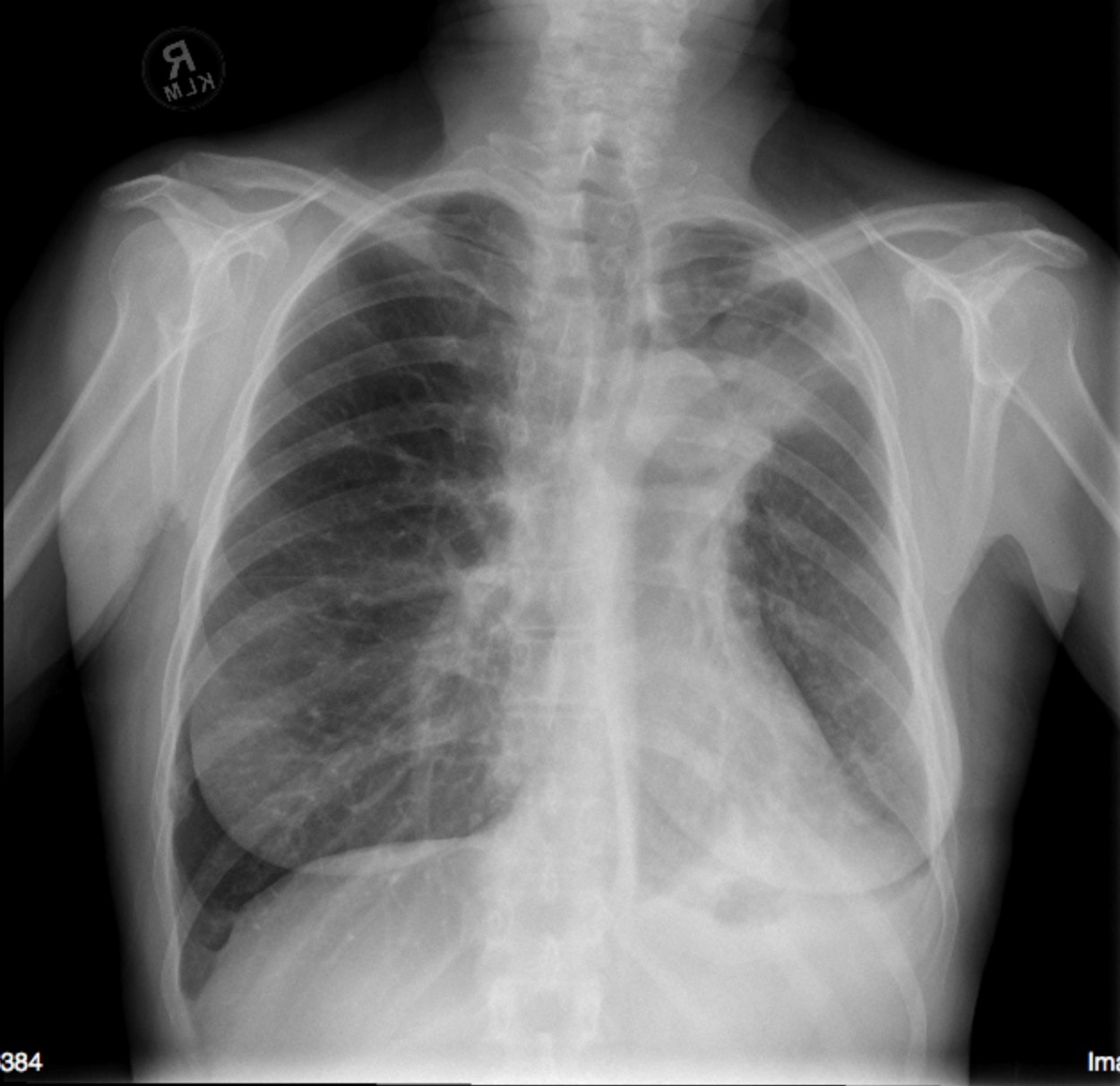
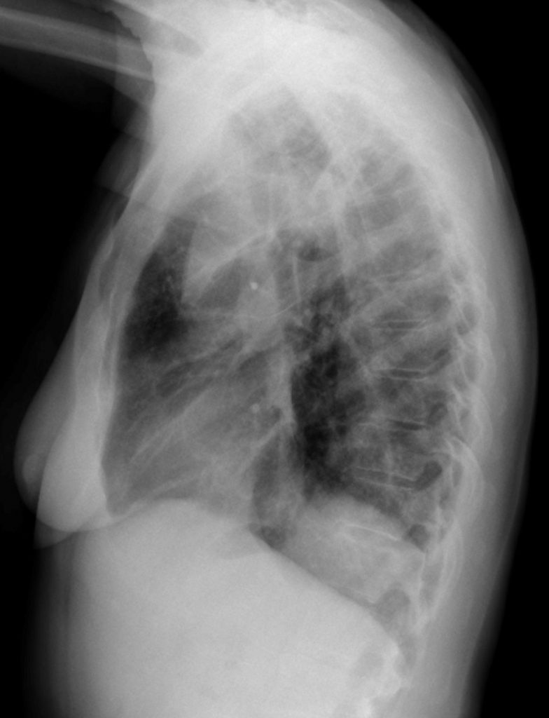
There was overall diminished volume in the left hemithorax (hemidiaphragm was elevated, the mediastinum was shifted to the left, there was vague haziness in left lung, and the trachea was deviated to the left). The left upper lobe was opaque and partially collapsed and there was a rounded margin of the hilar and left upper lung opacity suggesting an underlying tumor. The left upper lobe bronchus was surrounded by opacity and was irregular and constricted. The aortic shadow was obscured by the adjacent opaque lung. No obvious lymphadenopathy.
Interpretation:
Partial left upper lobe collapse with high suspicion for an associated left upper lobe lung mass. CT of the chest was recommended.
Diagnosis:
Left Upper Lobe Collapse
Pathology:
Adenocarcinoma of the Lung
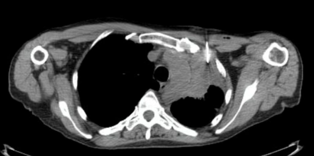
Limited CT (No contrast) for Core Needle Biopsy LUL Mass (Biopsy = Adenocarcinoma)

Discussion:
New and unexpected lobe or lung collapse is a very ominous finding in a patient over the age of fifty, especially if they have a history of cigarette smoking. If there are any imaging signs that suggest a mass lesion this requires immediate further investigation.
If the lung opacity is suspected to be related to community acquired pneumonia, with minimal imaging signs to suggest volume loss, the imaging abnormalities should initially be managed medically. If the radiographs do not improve with medical management i.e. complete resolution, the patient requires further investigation to determine if there is an underlying cause for the lung opacity .
Lung Malignancy
- Lung cancer is the most common fatal malignancy in men.
- Lung cancer is the second most common malignancy in women (next to breast cancer).
- The major sub-types of lung cancer are Adenocarcinoma, Squamous Cell Carcinoma, Small Cell Carcinoma and Large Cell.
- Primary lung cancer usually presents as a solitary nodule or mass. Adenocarcinoma often presents as a peripheral lung nodule or mass.
- More centrally situated lung malignancies may cause lobar or lung opacification with, or without, volume loss. A central, or hilar, location for lung malignancy is common with Small Cell Carcinoma.
X-ray findings may include:
- The collapsed lobe will be more opaque on imaging.
- Normally, fissures can be seen on x-rays when the fissure is in the same plane as the x-ray beam. It is seen as a thin white line composed of the visceral and parietal pleura of opposing lobes. The fissure abutting the collapsed lobe will migrate to a new location following the collapsed lobe.
- Often the fissure of the lung acts as a barrier to the spread of the lung opacity and there will be a sharp demarcation between the collapsed lung and the normal adjacent lung. The fissure will no longer be identifiable as a unique structure as it is no longer surrounded by aerated lung.
- The global volume of the hemithorax with the collapsed lobe will be diminished.
- This global volume loss may result in shifting of the mediastinum towards the collapsed lung.
- There may be an ipsilateral mass or hilar adenopathy visible.
- The bronchus supplying the collapsed lobe may not be visible.
Attributions
Figure 9.12A PA Chest x-ray of Left Upper Lobe Collapse by Dr. Brent Burbridge MD, FRCPC, University Medical Imaging Consultants, College of Medicine, University of Saskatchewan is used under a CC–BY-NC-SA 4.0 license.
Figure 9.12B Lateral Chest x-ray of Left Upper Lobe Collapse by Dr. Brent Burbridge MD, FRCPC, University Medical Imaging Consultants, College of Medicine, University of Saskatchewan is used under a CC–BY-NC-SA 4.0 license.
Figure 9.13 CT Core Needle Biopsy of Left Upper Lobe Mass by Dr. Brent Burbridge MD, FRCPC, University Medical Imaging Consultants, College of Medicine, University of Saskatchewan is used under a CC–BY-NC-SA 4.0 license.

