Chapter 10 – Gastrointestinal and Abdominal
Jaundice
ACR – Gastrointestinal – Jaundice
Case
Pancreatic Adenocarcinoma
Clinical:
History – Painless jaundice.
Symptoms – Icteric eyes and skin. Itchy.
Physical – The gallbladder was palpable and non-tender. There was icterus of the sclera and skin. The patient was pale and cachectic.
Laboratory – A pattern of obstructive liver enzymes was noted with elevated direct and indirect bilirubin.
DDx:
Choledocholithiasis
Biliary Tree Malignancy
Pancreatic Malignancy
Metastatic Disease
Imaging Recommendation
ACR – Gastrointestinal – Jaundice, Variant 2
CT of the Abdomen, with intravenous contrast


Imaging Assessment
Findings:
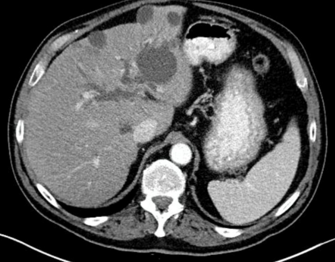
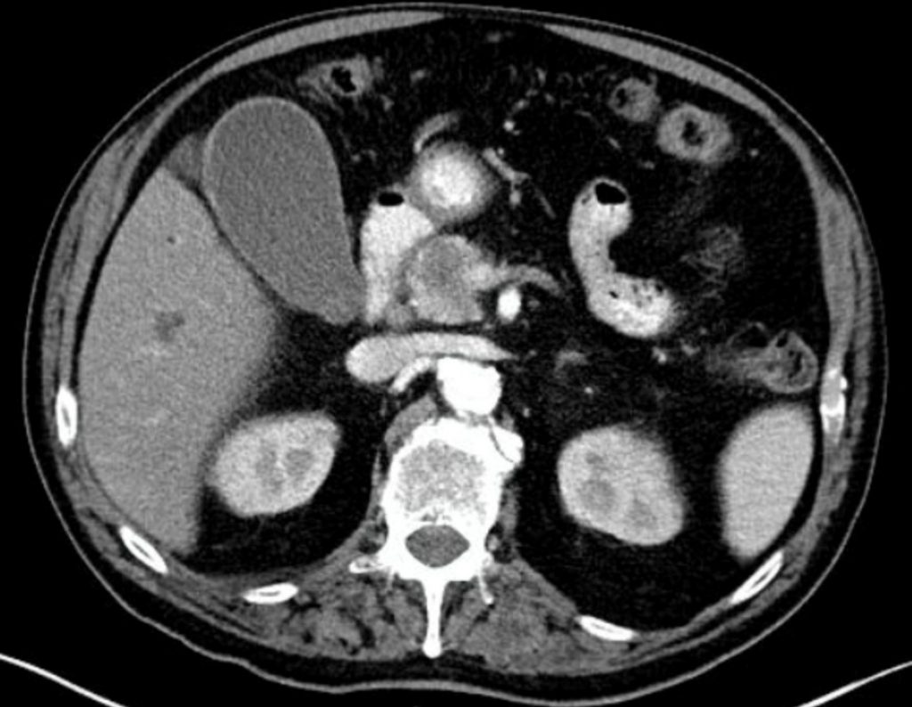
The contrast enhanced images demonstrated a non-enhancing mass in the head of the pancreas causing pancreatic duct and extrahepatic bile duct obstruction. There were multiple cysts in the liver. The intrahepatic bile ducts were dilated. No liver masses or nodules.
The pancreatic body and tail were very atrophic. No adenopathy.
Interpretation:
The findings were highly suggestive of a pancreatic malignancy.
Diagnosis:
Pancreatic Adenocarcinoma
Discussion:
One of the difficulties in determining a rational imaging strategy to evaluate jaundiced patients stems from the fact that jaundice is a clinical finding, not a single disease entity. The causes of non-hemolytic jaundice can be divided into two distinct categories: intrahepatic biliary stasis (hepatocellular jaundice) and mechanical biliary obstruction.
Because imaging plays little useful role in the evaluation of intrahepatic biliary stasis, the first task of the clinician caring for the jaundiced patient is to determine if jaundice is caused by bile duct obstruction. Several studies have shown that this distinction can be made in approximately 85% of patients using only clinical findings of age, nutritional status, pain, systemic symptoms, stigmata of liver disease, palpable liver, or gallbladder, and simple biochemical tests.
Patients with a high pretest probability of non-obstructive jaundice usually have either diffuse hepatocellular disease (e.g., cirrhosis, hepatitis), or, more rarely, inability of the liver to handle a bilirubin load (e.g., hemolytic anemia), or a metabolic deficiency (Gilbert’s disease). These patients need no imaging; liver biopsy is usually required.
Obstructive jaundice is jaundice resulting from obstruction to the flow of bile from the liver to the duodenum. In adults, extrahepatic (mechanical) obstruction accounts for 40% of patients presenting with jaundice as the primary symptom, and this likelihood increases with advancing age. The most common causes of obstructive jaundice are neoplasms of the pancreas, ampulla of Vater or biliary tract, choledocholithiasis, pancreatitis, and iatrogenic strictures of the biliary tree. Other less common causes include tumors metastatic to the biliary epithelium, sclerosing cholangitis, hepatic tumors adjacent to the liver hilum, perihepatic lymphadenopathy, and other causes of cholangitis.
Attributions
Figure 10.13A Axial CT of the liver demonstrating biliary system dilation by Dr. Brent Burbridge MD, FRCPC, University Medical Imaging Consultants, College of Medicine, University of Saskatchewan is used under a CC-BY-NC-SA 4.0 license.
Figure 10.13B Axial CT of the Pancreatic head demonstrating a hypodense mass in the head of the gland by Dr. Brent Burbridge MD, FRCPC, University Medical Imaging Consultants, College of Medicine, University of Saskatchewan is used under a CC-BY-NC-SA 4.0 license.

