Chapter 12 – Head and Neck
Facial Trauma
Case 1
Orbit Fracture
Clinical:
History – This child was struck in the left face by a soccer ball. She had left face pain and swelling.
Symptoms – The left eye was swollen shut.
Physical – No orbital globe injuries. Ocular movements were normal. No visual disturbance. The inferior orbital rim bone was tender.
DDx:
Facial bone fracture
Orbital Blow-out fracture
Maxillary sinus fracture
Imaging Recommendation
X-rays
Followed by CT if abnormal x-rays or abnormal eye muscle function.


Imaging Assessment
X-ray Findings:
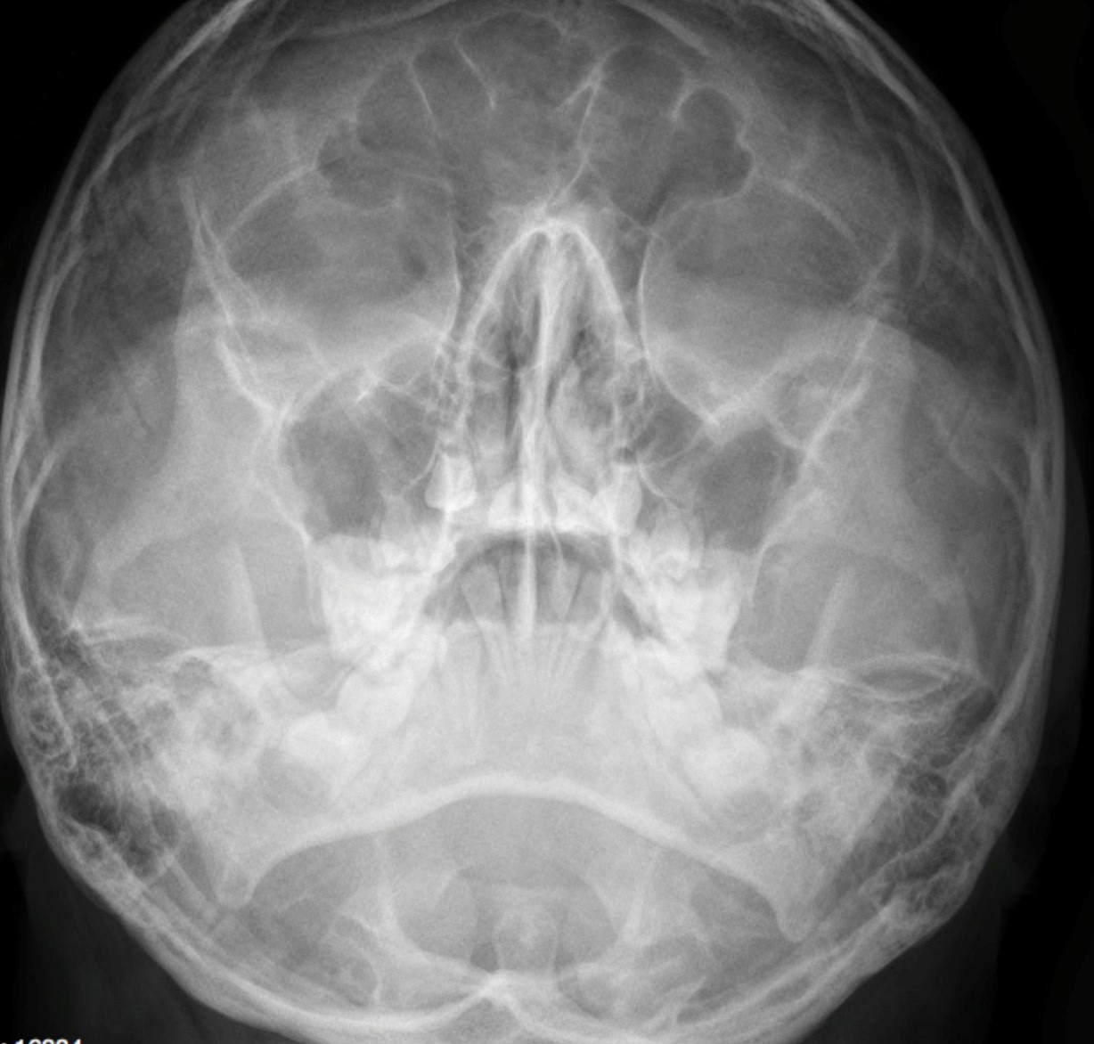
In the apex of the left maxillary antrum there was a very small, teardrop shaped, soft tissue, opacity. This opacity was only seen on the Water’s view. This was suggestive of injury of the apex of the maxillary antrum/floor of orbit. No obvious fractures seen.
Interpretation:
Possible floor of orbit fracture. CT of the orbits was recommended.
CT Findings:
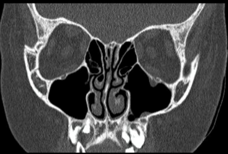
There was a depressed fracture of the floor of the left orbit/apex of the left maxillary sinus. This allowed a small amount of orbital fat to herniate through the bone defect. The ocular muscles were not herniating through the fracture. No hematoma or globe injury.
Diagnosis:
Teardrop Inferior Orbit – Left Floor of orbit fracture.
Discussion:
CT is the imaging study of choice for evaluating facial fractures. Multislice scanners allow for digital reconstruction in the sagittal and coronal planes so that the patient does not have to be repositioned in the scanner.
The most common orbital fracture is the blow-out fracture which is produced by a direct impact on the orbit (e.g., a baseball strikes the eye) and causes a sudden increase in intraorbital pressure leading to a fracture of the inferior orbital floor (into the maxillary sinus) or the medial wall of the orbit (into the ethmoid sinus). Sometimes the inferior rectus muscle can be trapped in the fracture, leading to restriction of upward gaze and diplopia.
Imaging findings of a blow-out fracture of the orbit may include:
- Orbital emphysema. Air in the orbit from communication with one of the adjacent air-containing, ethmoid, or maxillary sinus.
- Fracture through either the medial wall or floor of the orbit.
- Entrapment of fat and/or extraocular muscle, which projects downward as a soft tissue mass into the top of the maxillary sinus.
- Fluid (blood), causing an air – fluid level in the maxillary sinus.
Case 2
Complex Facial Fractures
Clinical:
History – This 25 year old male was kicked in the left face by a horse.
Symptoms – Pain in left face.
Physical – The face was swollen and painful. The mid-face was mobile on physical examination.
DDx:
Severe, Complex, Facial Bone Fractures
Imaging Recommendation
CT of the facial bones


Imaging Assessment
Findings:
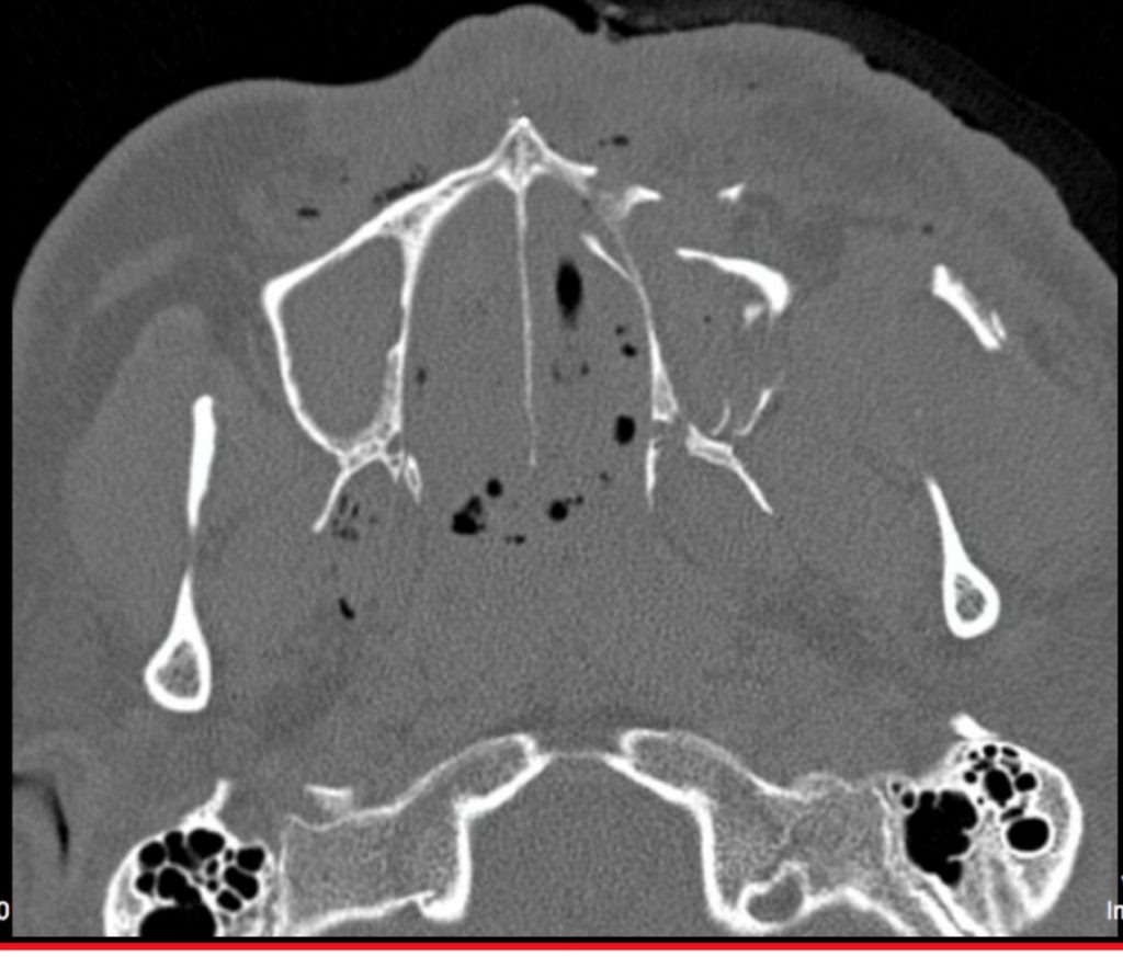
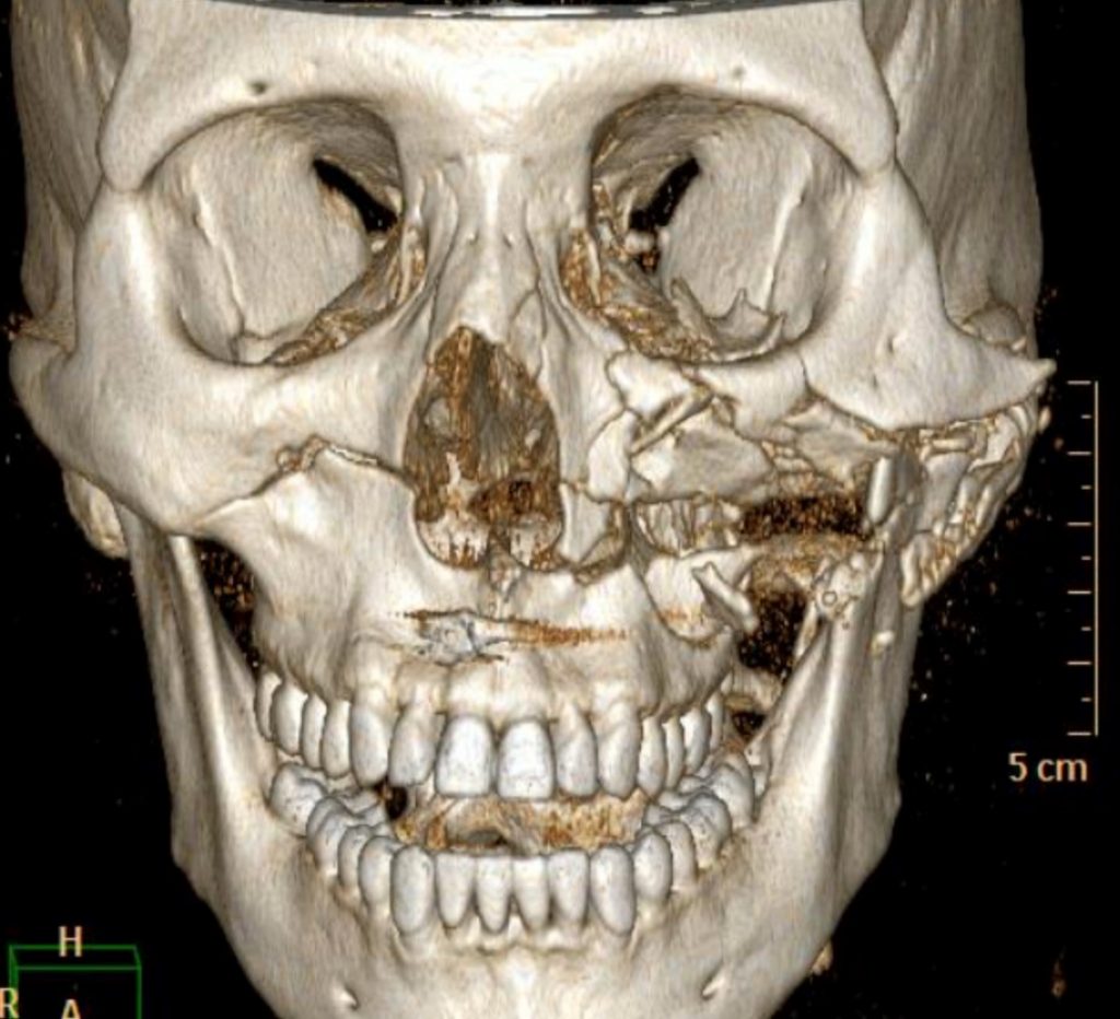
The left facial bones were pulverized including the maxillary bone, the zygoma, and the lateral wall of the orbit. This was a LeFort fracture type 3.
On the right side there is a LeFort fracture, type 2. Orbital contents were not herniating into the maxillary antrum. There was gas in the orbital space bilaterally.
Interpretation:
Complex Facial Bone Fractures.
Diagnosis:
Complex Facial Fractures – Trauma
Discussion:
CT is the imaging study of choice for evaluating facial fractures. Multislice scanners allow for digital reconstruction in the sagittal and coronal planes so that the patient does not have to be repositioned in the scanner.
Le Fort Facial Bone Fractures
Le Fort I fracture (horizontal), otherwise known as a floating palate, may result from a force of injury directed low on the maxillary alveolar rim, or upper dental row, in a downward direction. The key component of these fractures, in addition to pterygoid plate involvement, is involvement of the lateral bony margin of the nasal opening. They also involve the medial and lateral buttresses, or walls, of the maxillary sinus, traveling through the face just above the alveolar ridge of the upper dental row. At the midline, the inferior nasal septum is involved.
Le Fort II fracture (pyramidal) may result from a blow to the lower or mid maxilla. The key component of these fractures beyond the pterygoid plate fractures is involvement of inferior orbital rim. When viewed from the front, the fracture is classically shaped like a pyramid. It extends from the nasal bridge at or below the nasofrontal suture through the superior medial wall of the maxilla, inferolaterally through the lacrimal bones which contain the tear ducts, and inferior orbital floor through or near the infraorbital foramen.
Le Fort III fracture (transverse), otherwise known as craniofacial dissociation, may follow impact to the nasal bridge or upper maxilla. The salient feature of these fractures, beyond pterygoid plate involvement, is that they invariably involve the zygomatic arch, or cheek bone. These fractures begin at the nasofrontal and frontomaxillary sutures and extend posteriorly along the medial wall of the orbit, through the nasolacrimal groove and ethmoid air cells. The sphenoid is thickened posteriorly, limiting fracture extension into the optic canal. Instead, the fracture continues along the orbital floor and infraorbital fissure, continuing through the lateral orbital wall to the zygomaticofrontal junction and zygomatic arch. Within the nose, the fracture extends through the base of the perpendicular plate of the ethmoid air cells, the vomer, which are both part of the nasal septum. As with the other fractures, it also involves the junction of the pterygoids with the maxillary sinuses. CSF rhinorrhea, or leakage of the nutrient laden fluid that bathes the brain, is more commonly seen with these injuries due to ethmoid air cell disruption, as the air cells are located immediately beneath the skull base.
Attributions
Figure 12.2A Orbital x-ray demonstrating a teardrop opacity in the apex of the left maxillary antrum by Dr. Brent Burbridge MD, FRCPC, University Medical Imaging Consultants, College of Medicine, University of Saskatchewan is used under a CC-BY-NC-SA 4.0 license.
Figure 12.2B CT Scan of the Orbits with a teardrop opacity in the apex of the left maxillary antrum by Dr. Brent Burbridge MD, FRCPC, University Medical Imaging Consultants, College of Medicine, University of Saskatchewan is used under a CC-BY-NC-SA 4.0 license.
Figure 12.3A CT Scan of the Orbits and Facial region by Dr. Brent Burbridge MD, FRCPC, University Medical Imaging Consultants, College of Medicine, University of Saskatchewan is used under a CC-BY-NC-SA 4.0 license.
Figure 12.3B CT Scan 3D Rendering of Facial Bones by Dr. Brent Burbridge MD, FRCPC, University Medical Imaging Consultants, College of Medicine, University of Saskatchewan is used under a CC-BY-NC-SA 4.0 license.

