Chapter 9 – Chest
Emphysema
ACR – Chest – Chronic Dyspnea – Suspected Pulmonary Origin
Clinical:
History – Known history of chronic lung disease. The patient was a 50 pack/year cigarette smoker.
Symptoms – Cough – dry, shortness of breath, and wheezing.
Physical – Increased AP diameter, indrawing on inspiration, pursed lip breathing, distant breath sounds, hyperesonant chest percussion.
DDx:
Emphysema – Chronic Obstructive Pulmonary Disease (COPD)
Asthma
Imaging Recommendation
ACR – Chest – Chronic Dyspnea – Suspected Pulmonary Origin, Variant 1
Chest X-ray
Case 1
Moderate


Imaging Assessment
Findings:
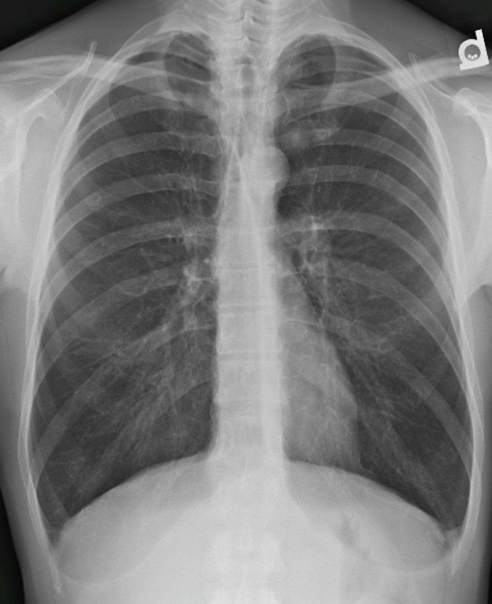
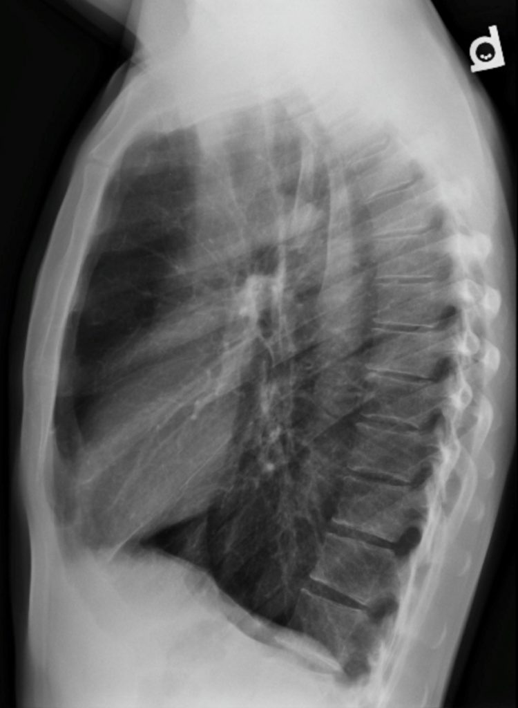
There was evidence of hyperinflation, including flattening of the diaphragm, especially on the lateral x-ray. The retrosternal clear space was widened and the AP diameter of the chest was increased. The lungs were hyperlucent with minimal deficiency of peripheral vascular markings. No evidence of pre-capillary hypertension.
Interpretation:
The abnormalities are in keeping with COPD.
Diagnosis:
COPD
Pathology:
Chronic Obstructive Pulmonary Disease
Discussion:
(COPD) is a long-term response to inhaled irritants or chemicals (tobacco smoke). This exposure leads to a cascade of inflammation, infection, and prostease enzyme imbalance that leads to the destruction of the connective tissues of the bronchi and acini of the lungs. The lungs become simplified into larger air containing spaces with damaged alveoli and bronchi.
The diagnosis of COPD is not made with chest radiographs but with clinical assessment and pulmonary ventilation testing to detect altered air flow related to expiratory obstruction during the breathing cycle. Radiographic findings may be very subtle to non-existent in the early stages of COPD.
- Chronic obstructive lung disease (COPD) is defined as a disease of airflow obstruction due to chronic bronchitis or emphysema.
- Chronic bronchitis is defined clinically by productive cough, whereas emphysema is defined pathologically by the presence of permanent and abnormal enlargement and destruction of the air spaces distal to the terminal bronchioles.
- Emphysema has three pathologic patterns
- Centriacinar (centrilobular) emphysema features focal destruction limited to the respiratory bronchioles and the central portions of the acinus. It is associated with cigarette smoking and is most severe in the upper lobes.
- Panacinar emphysema involves the entire alveolus distal to the terminal bronchiole. It is most severe in the lower lung zones and generally develops in patients with homozygous alpha 1 -antitrypsin deficiency.
- Paraseptal emphysema is the least common form. It involves distal airway structures, alveolar ducts, and sacs. Localized to fibrous septa or to the pleura, it can lead to formation of bullae, which may cause pneumothorax. It is not associated with airflow obstruction.
X-ray findings may include:
- Flattened hemidiaphragms due to overinflation of the lungs
- Increased AP diameter of the chest
- The retrosternal air space may become enlarged.
- The lungs become more lucent as the air spaces coalesce into larger simplified air containing regions.
- The vessels in the peripheral lung become cut-off and tapered.
- There may be large, air-containing, cystic spaces in the lungs (bulla)
Case 2
Severe
Clinical:
History – Known history of chronic lung disease. The 75 year old patient was a 60 pack/year cigarette smoker.
Symptoms – Cough – dry, shortness of breath, and wheezing. Unable to walk more than 50 steps due to severe shortness of breath.
Physical – Increased AP diameter, in drawing on inspiration, pursed lip breathing, distant breath sounds, hyperesonant chest percussion.
Imaging Recommendation
ACR – Chest – Chronic Dyspnea – Suspected Pulmonary Origin, Variant 1
Chest X-ray


Imaging Assessment
Findings:
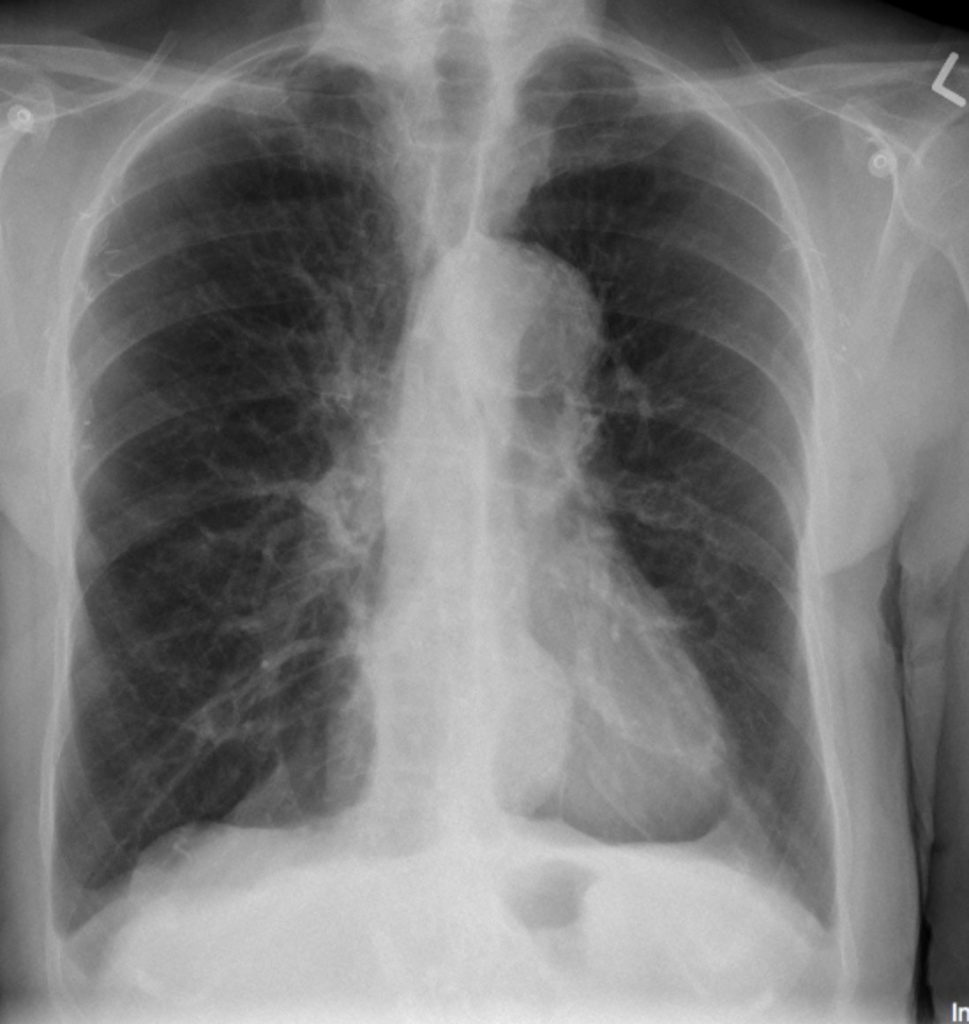
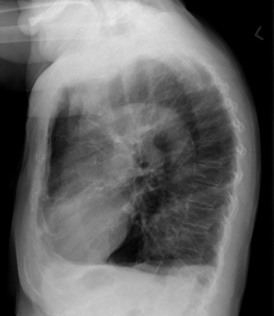
There was evidence of hyperinflation, including severe flattening of the diaphragm, especially on the lateral x-ray they were inverted. The retrosternal clear space was widened and the AP diameter of the chest was increased. The lungs were hyperlucent with a deficiency of peripheral vascular markings. There was enlargement of the central pulmonary arteries consistent with pre-capillary hypertension.
Interpretation:
The abnormalities are in keeping with COPD.
Diagnosis:
COPD – Severe, with pulmonary artery hypertension
Pathology:
Chronic Obstructive Pulmonary Disease
Attributions
Figure 9.22A PA Chest x-ray displaying moderate COPD by Dr. Brent Burbridge MD, FRCPC, University Medical Imaging Consultants, College of Medicine, University of Saskatchewan is used under a CC-BY-NC-SA 4.0 license.
Figure 9.22B Lateral Chest x-ray displaying moderate COPD by Dr. Brent Burbridge MD, FRCPC, University Medical Imaging Consultants, College of Medicine, University of Saskatchewan is used under a CC-BY-NC-SA 4.0 license.
Figure 9.23A PA Chest x-ray displaying severe COPD by Dr. Brent Burbridge MD, FRCPC, University Medical Imaging Consultants, College of Medicine, University of Saskatchewan is used under a CC-BY-NC-SA 4.0 license.
Figure 9.23B Lateral Chest x-ray displaying severe COPD by Dr. Brent Burbridge MD, FRCPC, University Medical Imaging Consultants, College of Medicine, University of Saskatchewan is used under a CC-BY-NC-SA 4.0 license.

