Chapter 10 – Gastrointestinal and Abdominal
Diverticulitis
ACR – Gastrointestinal – Left Lower Quadrant Pain
Case
Diverticulitis
Clinical:
History – 4 days of left lower quadrant pain.
Symptoms – The patient has had persistent, 6/10, pain in the left lower quadrant. Bowel function was normal.
Physical – Fever (40C). There was focal moderate tenderness in the left lower quadrant with deep palpation. Bowel sounds were normal. The rectal examination was normal.
Laboratory – The white blood cell count was elevated.
DDx:
Pelvic/Abdominal Abscess
Diverticulitis
Colitis
Enteritis
Imaging Recommendation
ACR – Gastrointestinal – Left Lower Quadrant Pain – Suspected Diverticulitis, Variant 1
CT Abdomen and Pelvis


Imaging Assessment
Findings:
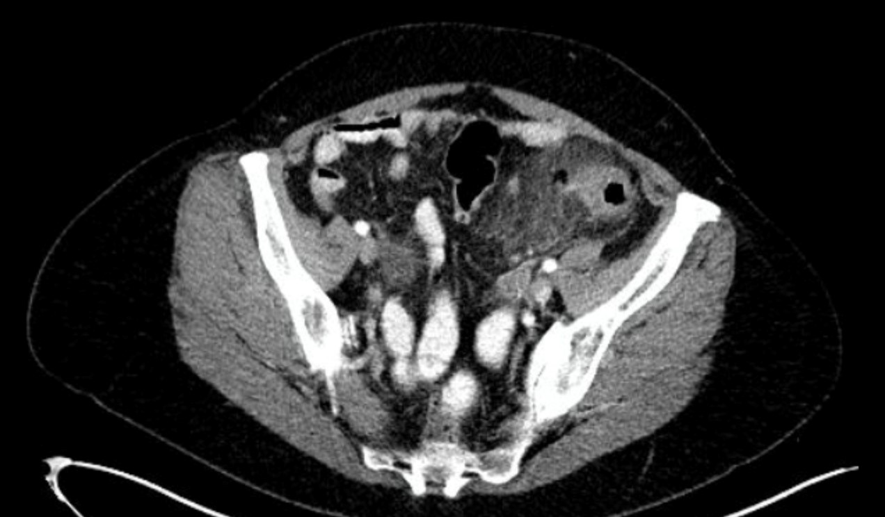
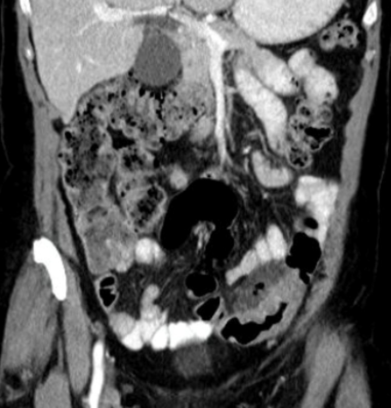
There was a very thick-walled segment of the sigmoid colon in the left lower quadrant. There were multiple diverticula in the region. The pericolonic fat was indurated. There was a long gas filled diverticulum that may represent a contained perforation of the colon.
Interpretation:
Diverticulitis with a possible contained perforation.
Diagnosis:
Diverticulitis
Colitis
Discussion:
Diverticula of the colon are herniations of mucosa and submucosa through the muscular layer of the colon. They are more common in the distal colon and increase in frequency with age.
At times they can become secondarily infected due to a partially obstructed or obstructed neck of the diverticulum. The diverticula contain fecal contents that can become infected within the closed space. CT is the optimal imaging modality for active diverticulitis.
CT findings of diverticulitis may include:
- Thickened colonic wall > 4 – 5 mm
- Pericolonic haziness due to inflammatory fluid/edema
- Larger fluid collections, abscesses or contained perforations
- Gas bubbles in the wall of the colon, the peri-colonic tissues, or in fluid adjacent to the colon.
- Pneumoperitoneum
The most common cause of left lower quadrant pain in adults is acute sigmoid and/or descending colon diverticulitis. It has been estimated that between 10% and 25% of patients with diverticulosis will ultimately develop diverticulitis
Triage for patients with suspected diverticulitis (i.e., left lower quadrant pain) should address 2 major clinical questions:
1) What are the differential diagnostic possibilities in this clinical situation?
2) What information is necessary to make a definitive management decision?
Some patients with acute diverticulitis may not require any imaging, notably those with typical symptoms of diverticulitis (e.g., left lower quadrant pain and tenderness) without suspected complications or those with a previous history of diverticulitis who present with clinical symptoms of recurrent disease. In some instances, such patients are treated medically without undergoing radiologic examinations, but diverticulitis can be simulated by other acute abdominal disorders.
Patients with diverticulitis may require surgery or interventional radiology procedures because of associated complications, including abscesses, fistulas, obstruction, or perforation. As a result, there has been a trend toward the greater use of medical imaging to confirm the diagnosis of diverticulitis, evaluate the extent of the disease, and detect complications before deciding on the most appropriate treatment.
Attributions
Figure 10.10A Axial CT of Abdomen by Dr. Brent Burbridge MD, FRCPC, University Medical Imaging Consultants, College of Medicine, University of Saskatchewan is used under a CC-BY-NC-SA 4.0 license.
Figure 10.10B Coronal CT of Abdomen by Dr. Brent Burbridge MD, FRCPC, University Medical Imaging Consultants, College of Medicine, University of Saskatchewan is used under a CC-BY-NC-SA 4.0 license.

