Chapter 14 – Musculoskeletal
Clavicle Fracture
ACR – MSK – Acute Shoulder Pain
Case
Clavicle fracture
Clinical:
History – This 32 year old male was checked into the boards during a hockey game. He had immediate pain in his left shoulder.
Symptoms – The patient had severe pain in his left shoulder region. It was difficult for him to move his arm due to pain. Range of motion of the gleno-humeral joint was mildly limited due to pain.
Physical – There was soft tissue swelling over the lateral clavicle and there was a hard protuberance in this region as well.
DDx:
Hematoma
Acromio-Clavicular joint dislocation
Clavicle fracture
Imaging Recommendation
ACR – MSK – Acute Shoulder Pain, Variant 1
Shoulder X-rays


Imaging Assessment
Findings:
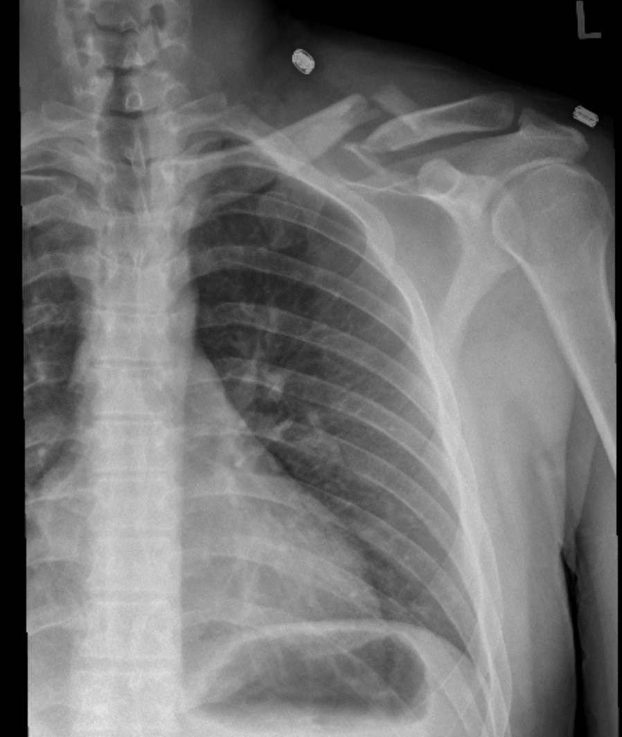
There was a comminuted, impacted, fracture of the left clavicle at the junction of the middle 1/3 and the lateral 1/3. The angle formed at the fracture site was mild to moderate and directed cranially. The angle created at the fracture site is due to the attachment of the sternocleidomastoid muscle pulling the medial fragment in a cranial direction.
Interpretation:
Impacted, comminuted, left clavicle fracture.
Diagnosis:
Clavicle fracture
Discussion:
Radiography is a useful initial screening modality for acute shoulder pain of all causes. Radiography is useful in the evaluation of fractures of the shoulder girdle. All radiographic shoulder studies should include frontal examinations. The frontal views can be straight antero-posterior projection (AP) with the humerus in neutral position or with the humerus in internal and/or without external rotation. Local protocols for radiographic evaluation of the shoulder for trauma vary widely. However, the shoulder trauma protocol should have at least three views, of which two views are orthogonal.
X-ray findings may include:
- The most common location for a clavicular fracture is at the junction of the lateral 1/3 and the middle 1/3.
- In children there may be an incomplete or greenstick type of fracture.
- Clavicle fractures may occur in the newborn with difficult deliveries.
- The medial clavicular fragment is typically cranially displaced due the pull of the sternocleidomastoid muscle.
- In most circumstances there is either a fracture of the clavicle or an acromio-clavicular joint dislocation and the two injuries are usually mutually exclusive.
Attributions
Figure 14.1A X-ray of the left shoulder, pre-operative, clavicle fracture by Dr. Brent Burbridge MD, FRCPC, University Medical Imaging Consultants, College of Medicine, University of Saskatchewan is used under a CC-BY-NC-SA 4.0 license.
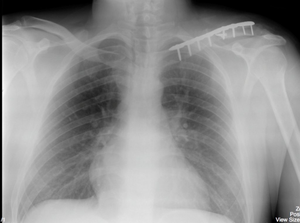
Figure 14.1B X-ray of the left shoulder, post-operative, clavicle fracture by Dr. Brent Burbridge MD, FRCPC, University Medical Imaging Consultants, College of Medicine, University of Saskatchewan is used under a CC-BY-NC-SA 4.0 license.

